
시각유발전위(visual evoked potential, VEP)는 섬광자극이나 모양역전(pattern reversal) 방법을 통해 시각적 자극을 주고, 유발된 전위를 후두부에서 측정하는 검사이다. 시각유발전위는 시각적 자극이 망막에서부터 시신경, 시신경교차(optic chiasm), 시각로(optic tract), 외측무릎체(lateral geniculate body), 시각부챗살(optic radiation) 및 일차시각피질(primary visual cortex)까지 도달하는 시각경로(visual pathway)의 기능을 평가할 수 있다[1]. 뇌하수체종양이나 두개인두종(craniopharyngioma)과 같이 시각경로와 인접한 구조물의 병변을 수술하는 경우, 시각경로의 손상이 발생할 위험이 높다. 따라서, 수술 중 시각경로의 손상 여부를 미리 확인하고, 시각 기능장애(visual dysfunction)를 예방하기 위해 시각유발전위를 이용하여 수술중신경계감시를 시행해 볼 수 있다. 저자들은 뇌하수체종양 환자에서 나비뼈경유수술(transsphenoidal surgery) 중 시각유발전위를 이용하여 시각경로의 기능을 효과적으로 감시하였던 증례를 보고하고, 수술중신경계감시에서 시각유발전위의 유용성에 대해 논의하고자 한다.
증례
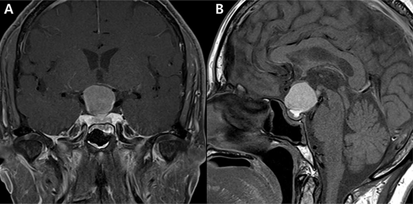
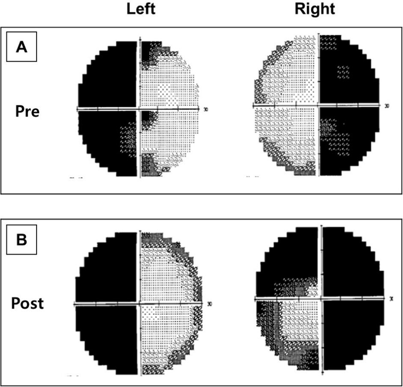
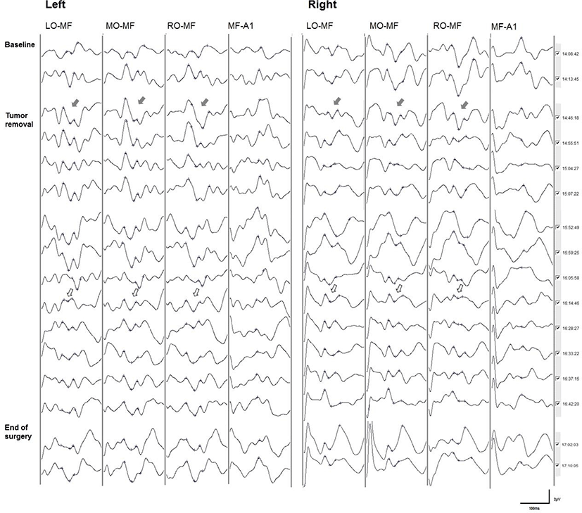
골다공증 외 특이 병력 없는 64세 여자가 내원 1주일 전부터 발생한 두통과 시야결손(visual field defect)으로 외래에 내원하였다. 신경학적 진찰 상 대면검사(confrontation test)에서 양귀쪽반맹(bitemporal hemianopsia) 소견이 확인되었으며, 그 외 신경학적 이상소견은 관찰되지 않았다. 뇌 자기공명영상검사에서 2.6 cm 크기의 덩어리가 안장/안장위 영역(sellar/suprasellar area)에서 관찰되었으며, 뇌하수체샘종(pituitary adenoma) 또는 라케트틈새낭(Rathke’s cleft cyst)이 의심되었다(Fig. 1). 상기 소견에 대하여 나비뼈경유수술(transsphenoidal surgery)을 통한 종양제거술을 시행하였다. 수술 전 험프리 시야검사(Humphrey perimetry)를 시행하였으며, 양귀쪽반맹(bitemporal hemianopsia)소견이 확인되었다(Fig. 2-A). 수술 전 시행한 시각유발전위에서는 양측 P100 잠복기(latency)는 모두 정상 범위로 확인되었다. 마취는 프로포폴(propofol) 4 µg/mL와 레미펜타닐(remifentanil) 3 ng/mL를 사용하여 완전정맥마취(total intravenous anesthesia, TIVA)를 시행하였다. 수술중신경계감시는 뇌파, 체성감각유발전위 및 시각유발전위를 시행하였으며, Cadwell Cascade Elite(Cadwell Industries, Inc., Washington, USA) 장비를 이용하여 검사하였다. 체성감각유발전위는 상지에서는 정중신경(median nerve)에서 자극하고, C3’-Fz, C4’-Fz에서 측정하였으며, 하지에서는 뒤경골신경(posterior tibial nerve)에서 자극하고, Cz’-Fz 전극에서 측정하였다. 자극 빈도는 2.82 Hz로 시행하였고, 평균화 횟수는 200번으로 하였다. 뇌파는 C3, C4, Fz, Cz, LO, MO, RO, MF 전극을 이용하여 측정하였다. 시각유발전위는 고글을 이용하여 섬광자극을 주었으며, LO, MO, RO, A1-MF의 4채널 전극에서 기록하였다. 섬광자극의 빈도는 1.05 Hz이며, 주파수 폭(Band-pass)은 High-pass filter는 70 Hz, Low-pass filter는 1 Hz로 설정하고, 평균화 횟수는 100번으로 설정하였다. 체성감각유발전위 및 시각유발전위의 진폭이 50% 이상 감소하는 경우를 유의한 변화로 간주하였다. 전체 수술 기간 동안 뇌파와 체성감각유발전위에서 유의한 변화는 관찰되지 않았다. 시각유발전위는 수술 시작 시 정상적으로 파형이 형성되었으나, 종양제거술을 시작하면서 양측 시각유발전위의 진폭(amplitude)이 감소하였다가 회복되기를 반복하였다. 종양제거술 중 양측 시각유발전위의 유의한 진폭 감소가 확인되었으며, 좌측에 비해 우측에서 진폭의 감소가 현저하였다(Fig. 3). 감소된 시각유발전위의 진폭은 수술 종료 시까지 회복되지 않았다. 수술 후 시행한 시각유발전위에서 우측 P100 잠복기가 지연된 소견이 관찰되었으며, 시야검사에서는 우안의 시야장애가 악화된 소견이 관찰되었다(Fig. 2-B).
고찰
시각 기능은 환자의 삶의 질에 미치는 영향이 크기 때문에 시각경로에 인접한 병변을 수술하는 경우에는 시각 기능을 보존 및 시각 기능장애를 예방하는 것이 중요하다. 신경외과수술 중 시각경로에 인접하여 발생한 뇌하수체선종(pituitary adenoma), 두개인두종, 교차신경교종(chiasmal glioma), 후두엽교종(occipital lobe glioma) 등 종양절제술이나 내경동맥 clinoid 동맥류 clipping 등은 수술 후 시각 기능장애가 발생할 위험성이 높은 수술이다. 따라서 이러한 수술을 시행하는 경우, 시각경로에 대한 수술중신경계감시를 시행하는 것이 필요하다. 시각유발전위는 시각적 자극에 의해 유발된 전위를 후두엽에서 측정하는 검사로, 망막에서부터 일차시각피질까지 시각경로를 평가할 수 있어 수술 중 시각경로의 이상을 감시하는데 활용할 수 있다.
수술 중 시각유발전위는 1973년에 Wright 등이 안와종양(orbital tumor) 수술에서 처음으로 시행하였으며, 수술 중 시각경로 손상 여부를 감시하고, 수술 후 시각 기능장애를 예방하는데 유용한 검사로 생각되었다. 하지만, 일부 연구에서는 시각유발전위의 재현성과 신뢰도가 낮고, 시각유발전위의 변화와 수술 후 임상적 예후의 연관성이 없어 임상적으로 유용하지 않다고 주장하였다[2,3]. 이러한 결과는 마취제의 종류, 섬광자극의 방법 및 자극강도 등이 영향을 미쳤을 것으로 생각된다. 하지만, 최근에는 마취제의 발달로 완전정맥마취가 가능해지고, 광선자극 장치의 발달하면서 보다 안정적인 시각유발전위를 획득할 수 있게 되었다. 일부 연구에서는 고강도 발광 다이오드를 이용하여 시각유발전위를 시행한 결과, VEP의 재현성이 93% 이상으로 높아졌으며, 수술 중 시각유발전위의 관찰된 유의미한 변화(50% 이상 진폭감소 또는 완전 소실)를 보인 경우에는 수술 후 시야결손이 악화되는 소견을 보인 결과도 보고되었다[4,5].
시각유발전위는 일반적으로 모양역전(pattern reversal) 방법 또는 섬광자극(flash stimulation)을 이용하여 망막에 시각적 자극을 줄 수 있지만, 전신마취 상태에서는 섬광자극을 통해서만 검사를 시행할 수 있다. 섬광자극을 주는 방법은 안정적인 시각유발전위를 획득하는데 중요한 요인 중 하나이다. 다양한 방법의 섬광자극이 사용되었으며, 최근에는 고강도 발광 다이오드(high- intensity light emitting diode)를 이용한다. 섬광자극 주기는 4 Hz 이하로 자극을 주며, 일반적으로 약 1–2 Hz 주기로 자극을 주게 된다[1]. 섬광자극 주기가 높아질수록 시각유발전위의 진폭은 점차 감소되어 파형을 얻기 어려워진다. 시각유발전위를 실시 할 때 망막전위도검사(electroretinography)를 이용하면 자극이 망막에 도달하는지 확인하는데 도움이 된다[4,5]. 시각유발전위는 75 msec 주변에서 관찰되는 maximal negative wave(N75)와 100 ms 주변에서 관찰되는 maximal positive wave(P100) 사이의 peak-to-peak 진폭을 시각유발전위의 진폭으로 정의한다[6]. 시각유발전위의 경보 기준은 진폭이 50% 이상 감소되는 경우는 유의한 변화로 간주한다[4,5]. 일부 연구자에서는 20% 이상 진폭 감소를 의미 있는 변화로 간주하기도 한다[7]. 시각유발전위가 완전 소실되는 경우에는 수술 후 심한 시각 기능장애가 발생할 가능성이 높다[5].
시각유발전위는 다양한 요인들에 의해 영향을 받는다. 마취제, 수술 전 시각 기능 상태, 체온, 혈압 등이 영향을 미칠 수 있으며, 특히 마취제는 시각유발전위에 매우 강한 영향을 미친다. 마취제 중 모든 흡입마취제는 시각유발전위를 억제시키며, 마취제의 농도가 높을수록 시각유발전위의 잠복기가 연장되고 및 진폭의 감소 정도가 더 커진다[8]. 정맥마취제 중 프로포폴(propofop), 펜타닐(fentanyl)이나 레미펜타닐(remifentanil)은 VEP에 미치는 영향이 적은 편이다[9]. 따라서 수술 중 시각유발전위를 시행하는 경우 프로포폴이나 레미펜타닐을 이용하여 완전정맥마취를 시행하는 것이 좋다. 하지만 고용량의 프로포폴을 사용하는 경우에는 시각유발전위의 감쇠(attenuation)가 관찰될 수 있다. 근육이완제는 시각유발전위에 영향을 미치지 않는다. 수술 전 환자의 시각 기능장애가 심한 경우에는 시각유발전위의 재현성이 낮고, 파형을 기록하기 어려울 수 있다. 환자가 실명 상태이거나 심한 시야결손이 있는 경우, 충분한 시각적 자극이 이뤄지지 않아 시각유발전위의 파형을 획득하기 어려울 수 있다. 체온도 시각유발전위에 영항을 미치는 요인 중 하나이다. 수술 중 체온이 낮아지면 시각유발전위의 진폭은 감소하고 잠복기는 연장되는 소견을 보인다. 체온이 25℃–27℃로 낮아지면 시각유발전위의 파형은 소실된다[10]. 이외에도 저탄산혈증, 저산소증, 저혈압, 혈액희석(hemodilution)도 시각유발전위의 파형에 영향을 미치는 것으로 알려져 있다.
본 증례에서는 완전정맥마취 하에 섬광자극 시각유발전위를 이용하여 나비뼈경유수술 중 시신경 기능을 실시간으로 감시하였다. 특히 본 증례에서는 종양제거술 중 시각유발전위의 진폭이 감소하는 소견이 관찰되었고, 좌측보다는 우측에서 진폭의 감소가 더 큰 양상을 보였다. 이에 우측 시신경의 기능 이상이 의심되었으며, 감소된 진폭은 수술 종료 시까지 회복되지 않아 수술 후 우측 시신경 기능의 이상이 발생할 가능성이 높을 것으로 예상되었다. 수술 후 시행한 시야검사에서 우측 내측 시야장애가 새로이 발견되었는데, 이는 시각유발전위의 유의미한 변화가 수술 후 임상적 예후와 연관이 있음을 보여준다. 또한, 본 증례에서 시행한 나비뼈경유수술은 내시경비내접근법(endoscopic endonasal approach)을 통해 종양을 제거함으로써 절개부위가 작고, 조직손상이 적어 수술 후 합병증이 적다는 장점이 있지만, 시신경교차부위의 병변을 제거할 때 시신경의 손상을 초래할 위험성이 있다. 그러므로 나비뼈경유수술을 시행할 때 시각유발전위를 통해 수술 중 시각 기능을 실시간으로 감시하고, 수술 후 시각 기능장애를 예측하는데 도움을 받을 수 있다.
수술 중 시각유발전위감시는 과거 연구들에서 임상적 유용성이 명확하지 않았다고 알려졌으나, 최근 연구에서는 검사의 재현성이 높아지면서 시각 기능을 보존하고, 수술 후 시각 기능장애를 예측하는데 유용한 검사로 여겨지고 있다. 앞서 살펴본 바와 같이 다양한 요인들이 시각유발전위에 영향을 줄 수 있으므로 주의가 필요하며, 적절한 자극, 마취방법 그리고 다양한 요인들에 대한 고려하여 검사한다면 시각 기능장애가 발생할 위험성이 높은 수술에서 매우 유용하게 이용될 수 있을 것이다.