Journal of Intraoperative Neurophysiology
Korean Society of Intraoperative Neurophysiological monitoring
Case Report

미주신경초종 수술 중 성대 근전도 감시의 유용성: 증례보고

안상일1https://orcid.org/0000-0002-2416-6174, 정하늘1,*https://orcid.org/0000-0002-5785-116X
Sang-Il Ahn1https://orcid.org/0000-0002-2416-6174, Ha-Neul Jeong1,*https://orcid.org/0000-0002-5785-116X
1연세대학교 의과대학 세브란스병원 신경과
1Department of Neurology, Severance Hospital, Yonsei University College of Medicine, Seoul, Korea
*Corresponding author: Ha-Neul Jeong, Department of Neurology, Severance Hospital, Yonsei University College of Medicine, 50-1, Yonsei-ro, Seodaemun-gu, Seoul 03722, Korea, Tel: +82-2-2008-5260, E-mail: hnjeong@yuhs.ac

© Copyright 2019 Korean Society of Intraoperative Neurophysiological Monitoring. This is an Open-Access article distributed under the terms of the Creative Commons Attribution Non-Commercial License (http://creativecommons.org/licenses/by-nc/4.0/) which permits unrestricted non-commercial use, distribution, and reproduction in any medium, provided the original work is properly cited.

Received: Jun 29, 2019 ; Revised: Jul 29, 2019 ; Accepted: Aug 17, 2019

Published Online: Sep 30, 2019

ABSTRACT

Postoperative vagal dysfunction is a serious complication can lead to dysphonia, dysphagia, pharyngeal or airway obstruction. Therefore, intraoperative monitoring of vagus nerve during surgeries involving the lower brain stem is important to prevent such deficits. Monitoring of vagus nerve can be performed by monitoring free running and stimulated electromyography from the vocal cords. Recording using adhesive surface electrodes on the endotracheal tube approximating the vocal cords can be used to monitor vagus nerve. We report a case of vagal schwannoma with intraoperative vocal cord electromyographic monitoring during the craniotomy and tumor removal. Our case suggests that intraoperative vagus nerve monitoring using surface electrodes on the endotracheal tube could be aid in identifying neural structures and assessing the functional integrity.

Keywords: intraoperative neurophysiological monitoring; vagus nerve; vocal cord

서론

미주신경(vagus nerve)은 연수에서 시작하여 인두, 성대, 심장, 내장기관 등에 이르는 긴 신경이며, 운동 신경, 감각 신경, 자율 신경을 포함하고 있는 복잡한 혼합신경이다. 미주신경이 손상된 경우 연하곤란, 발성장애, 호흡곤란을 초래하게 되며, 심한 경우에는 기관창냄술이나 위창냄술을 시행하게 되는 등 환자의 삶의 질에 미치는 영향이 치명적이다[1]. 따라서 미주신경 손상의 가능성이 높은 뇌 기저나 아래 뇌간 부위의 수술 중에는 이를 예방하려는 노력이 필요하다. 이에 수술 중 미주신경의 신경생리학적 감시는 신경의 위치를 확인하고, 기능을 평가하기 위한 방법이다[2].

수술 중 미주신경을 감시하기 위해서는 인후두 부위의 근육에 대한 근전도검사를 이용하는데, 표면 전극이 부착된 기관내 튜브(endotracheal tube)를 이용하여 성대 근전도(vocal cord electromyography)를 기록하는 방법을 사용하기도 한다[3]. 이 비침습적인 방법은 갑상선 수술에서 미주신경의 가지인 되돌이후두신경(recurrent laryngeal nerve)을 보존하기 위한 방법으로 유용하게 사용되고 있다[4]. 본 논문에서는 미주신경 신경초종(schwannoma)을 제거하기 위한 개두술에서 수술 중 미주신경 감시를 위해 성대 근전도검사를 시행한 증례를 보고하고자 한다.

증례

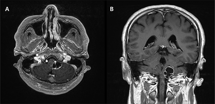
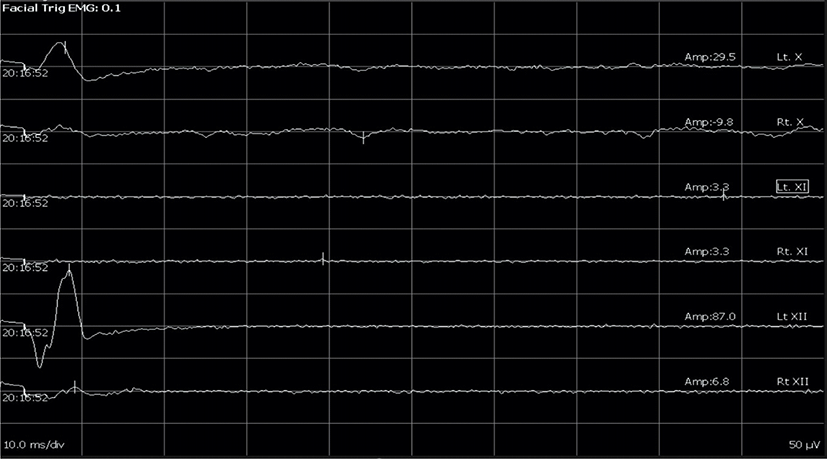
2012년 요추 신경초종으로 수술 받은 후 특별한 증상없이 지내던 47세 남자 환자가 어지럼증과 보행 시 한 쪽으로 기우는 증상으로 타 병원에서 시행한 검사상 뇌종양이 의심되어 수술적 치료를 받기 위해 본원으로 입원하였다. 입원 후 시행한 문진 상 좌측 청력 저하와 간헐적으로 사레가 들리는 증상을 호소하였다. 뇌 자기공명영상검사 상 좌측 소뇌다리뇌각 부위에 조영 증강되는 2.5 cm 크기의 뇌신경 신경초종이 의심되는 병변이 관찰되었다(Fig. 1). 환자는 뇌종양 제거를 위한 개두술을 받게 되었고, 마취는 프로포폴(propofol)과 레미펜타닐(remifentanil)을 이용한 완전정맥마취(total intravenous anesthesia) 방법을 이용하였다. 근이완제는 기관내삽관을 시행하기 위해 로쿠로늄브롬화물(rocuronium bromide)을 1회 정맥주사하였고, 이후 수술 과정에서는 사용하지 않았다. 수술 중 신경계 감시를 위하여 NIM-ECLIPSE System® (Medtronic Xomed Inc., Jacksonville, USA) 장비를 사용하였다. 사용한 기관내 튜브(NIM EMG Endotracheal Tube 6.0 mm I.D. × 8.8 mm O.D. [Medtronic Xomed Inc., Jacksonville, USA])에는 수술 중 미주신경 감시를 위해 성대 근전도 파형을 기록하기 위한 표면 전극이 부착되어 있었다. 튜브 위의 표면 전극들은 양측으로 한 쌍씩 부착되어 있어서 활성 전극과 참고 전극의 전위차를 기록할 수 있게 설정되어 있었다. 마취과 의사는 기관내삽관 시 표면 전극이 성대 부위에 위치하도록 주의하였다. 미주신경 감시와 더불어 11, 12번 뇌신경을 감시하기 위해서 양측 승모근(trapezius)과 혀(tongue) 근육에도 근전도 검사를 위한 바늘을 각각 한 쌍씩 삽입하였다. 접지 전극은 엉덩이 부위에 부착하였다. 기록 전극들의 임피던스(impedance)는 수술 종료 시까지 5 kOhms 이하로 유지하였다. 주파수 폭(band-pass)은 High-pass filter 1,500 Hz, Low-pass filter 30 Hz으로 설정하였고, Gain과 Sweep speed는 각각 50 µV/div, 10 ms/div로 하였다. 유발 근전도검사를 위해서 Concentric bipolar stimulator를 이용하여 종양 주변의 뇌신경이 있을 것으로 생각되는 부위에 자극하였다. 자극의 빈도, 세기, 지속시간은 각각 5 Hz, 0.1 mA, 200 µs으로 하였다. 자극에 의한 복합근육활동전위(compound muscle action potential)가 유발된 경우, 복합근육활동전위가 관찰된 근육들을 바로 신경외과 의사에게 알렸다((Fig. 2). 신경외과 의사는 종양의 아래 부위에 붙어있는 척추 동맥(vertebral artery)과 10-12번 뇌신경의 잔뿌리(rootlet)들을 박리하였고, 그 중 10번 신경 잔뿌리가 종양의 근원인 것으로 확인되었다. 수술 중 신경계 감시를 통해 신경외과 의사는 신경의 위치를 확인하여 신경 손상을 최소화하면서 종양을 제거하였으며, 전기 자극을 통해 미주신경의 정상적인 기능을 확인하였다. 수술 소견과 조직검사를 통합하여 좌측 미주신경의 신경초종으로 최종 진단되었으며, 수술 후 시행한 뇌 자기공명영상검사에서 종양은 완전 제거되었음을 확인하였다. 수술 후 2개월 추적 관찰 시까지 연하 장애나 발성 장애는 관찰되지 않았다.

join-1-2-35-g1
Fig. 1. T1 weighted brain magnetic resonance imaging with gadolinium enhancement (axial [A] and coronal view [B]) showed a 2.5 cm sized mass (white arrowhead) at the left cerebellopontine angle. It was presumed to be a cystic schwannoma originated from lower cranial nerve.
Download Original Figure
join-1-2-35-g2
Fig. 2. Bilateral vagus, glossopharyngeal and hypoglossal nerves were monitored by electromyogram of the vocal cord, trapezius, and tongue muscles, respectively. During the resection of a vagal schwannoma, left cranial nerve 10 and 12 were identified using the evoked compound muscle action potentials by stimulation around the tumor.
Download Original Figure

고찰

수술 중 아래 뇌신경(lower cranial nerve) 감시는 주로 뇌 기저와 하부 뇌간 부위의 수술에서 유용하며 근전도검사로 시행되는데, 신경 손상을 실시간으로 확인하기 위한 추적 근전도와 신경의 위치 확인과 기능 평가를 위한 유발 근전도의 두 가지 방법이 있다[3,5]. 혀인두신경(glossopharyngeal nerve)은 붓인두근(stylopharyngeus), 미주신경은 인후두 근육들(laryngeal and pharyngeal muscles), 더부신경(accessory nerve)은 목빗근(sternocleidomastoid)과 승모근(trapezius), 혀밑신경(hypoglossal nerve)은 혀(tongue) 근육을 이용하여 검사를 시행한다[6]. 그러나 붓인두근은 인후두 근육과 인접하게 위치하여 10번 뇌신경에 의한 근전도 파형이 관찰될 수 있기 때문에 9번 뇌신경 감시가 필요한 경우에는 10번 뇌신경 감시를 함께 시행하는 것이 권고된다[3].

아래 뇌신경 중 9, 10번 뇌신경 감시를 위한 인후부의 근육들은 정확하게 바늘 전극을 꽂는 것이 어렵고, 바늘에 의한 혈종의 우려가 있어서 비 침습적 방법인 표면 전극을 이용하여 기록하는 방법이 사용되기도 한다. 혀인두신경 감시에는 후두의 마스크 기도 유지기(laryngeal mask airway) 위의 표면 전극을 이용한 증례[7]가 보고된 바 있다. 미주신경 감시의 비 침습적인 방법으로는 기관내 튜브의 표면 전극을 이용한 성대 근육의 근전도검사를 시행한다. 고려해야 할 점은 기록 전극이 성대 부위에 위치되었는지 확인하는 것이며, 이를 위해서 후두경을 이용하는 것이 권고된다. 또한 미주신경은 폐와 심장을 비롯한 내장 기관의 부교감 기능을 담당하기 때문에 유발 근전도검사를 위한 신경 자극 시에 발생할 수 있는 부작용을 대비해야 한다는 것이다. 따라서 마취과 의사의 협조가 필요하며, 미주신경 자극에 의한 서맥과 저혈압에 대비하여 아트로핀(atropine)을 준비해야 한다[3]. 기관내 튜브의 표면 전극을 이용한 미주신경의 성대 근전도검사는 갑상선 수술의 합병증인 되돌이후두신경 손상을 예방하는 데 임상적 유용성이 입증되어 보편적으로 사용되고 있다[4]. 반면, 두개 내 수술에서 성대 근전도검사는 보고된 바가 적은 편이다.

본 증례에서는 개두술을 통한 좌측 미주신경초종의 제거 수술에서 성대 근전도검사를 이용한 미주신경 감시의 유용성을 확인하였다. 본 증례와 같이 미주신경초종의 수술에서 근전도검사를 이용한 수술 중 감시가 최근 보고되고 있다[1]. 신경초종의 경우에는 종양에 의해 정상 신경다발이 밀려나거나 구조가 변형되기 때문에 종양 제거 도중 정상 신경다발이 손상될 우려가 있다. 따라서 정상 신경의 위치를 확인하기 위한 유발 근전도검사는 신경초종의 수술에서 유용하게 사용될 것으로 생각된다. 실제로 전정신경초종(vestibular schwannoma) 제거 수술에서는 근전도검사를 이용한 안면신경 감시의 임상적 이점에 대해서 반론이 없으며, 적극적으로 사용되고 있다[8]. 추후 두개 내 수술에서 성대 근전도검사가 활성화되면 미주신경초종 수술에서의 유용성도 확인할 수 있을 것이다.

결론적으로, 기관내 튜브의 표면 전극을 이용한 성대 근전도검사를 이용한 수술 중 미주신경 감시는 뇌 바닥 혹은 하위 뇌간 부위의 두개내 수술에서 미주신경의 위치를 확인하고 기능을 평가하는 데 도움이 될 것으로 생각된다. 추후 임상적 예후를 예측하기 위한 근전도검사의 해석에 대해서 추가적인 연구가 필요할 것으로 생각된다.

Notes

Ethical approval

This article does not require IRB/IACUC approval because there are no human and animal participants.

Notes

Conflicts of interest

No potential conflict of interest relevant to this article was reported.

References

1.

Sandler ML, Sims JR, Sinclair C, Sharif KF, Ho R, Yue LE, et al. Vagal schwannomas of the head and neck: a comprehensive review and a novel approach to preserving vocal cord innervation and function. Head Neck. 2019;41(7):2450-66.

2.

Topsakal C, Al-Mefty O, Bulsara KR, Williford VS. Intraoperative monitoring of lower cranial nerves in skull base surgery: technical report and review of 123 monitored cases. Neurosurg Rev. 2008;31(1):45-53.

3.

Singh R, Husain AM. Neurophysiologic intraoperative monitoring of the glossopharyngeal and vagus nerves. J Clin Neurophysiol. 2011;28(6):582-6.

4.

Randolph GW, Dralle H, Abdullah H, Barczynski M, Bellantone R, Brauckhoff M, et al. Electrophysiologic recurrent laryngeal nerve monitoring during thyroid and parathyroid surgery: international standards guideline statement. Laryngoscope. 2011;121 Suppl 1:S1-16.

5.

Skinner SA. Neurophysiologic monitoring of the spinal accessory nerve, hypoglossal nerve, and the spinomedullary region. J Clin Neurophysiol. 2011;28(6): 587-98.

6.

Husain AM. A practical approach to neurophysiologic intraoperative monitoring. New York, NY: Demos Medical; 2008. p. 23.

7.

Husain AM, Wright DR, Stolp BW, Friedman AH, Keifer JC. Neurophysiological intraoperative monitoring of the glossopharyngeal nerve: technical case report. Neurosurgery. 2008;63 Suppl 2:277-8.

8.

Youssef AS, Downes AE. Intraoperative neurophysiological monitoring in vestibular schwannoma surgery: advances and clinical implications. Neurosurg Focus. 2009;27(4):E9.