Case Report
경부척수증 수술 중 자세를 잡는 과정에서의 신경계감시: 증례 보고
Nervous system monitoring in the positioning process of cervical myelopathy surgery: a case report
Namo Jeon
1
, Hyeong-Wook Han
1, Soo-bin Lee
2, Doo Young Kim
1,*
1가톨릭관동대학교 의과대학 국제성모병원 재활의학과
2가톨릭관동대학교 의과대학 국제성모병원 정형외과
1Department of Rehabilitation Medicine, International St Mary’s Hospital, Catholic Kwandong University College of Medicine, Incheon, Korea
2Department of Orthopaedic Surgery, International St Mary’s Hospital, Catholic Kwandong University College of Medicine, Incheon, Korea
*Corresponding author: Doo Young Kim, Department of Rehabilitation Medicine, International St Mary’s Hospital, Catholic Kwandong University College of Medicine, Incheon 22711, Korea, Tel: +82-32-290-3112, Fax: +82-32-290-3879, E-mail:
[email protected]
© Copyright 2023 Korean Society of Intraoperative Neurophysiological Monitoring. This is an Open-Access article distributed under the terms of the
Creative Commons Attribution Non-Commercial License (http://creativecommons.org/licenses/by-nc/4.0/) which permits
unrestricted non-commercial use, distribution, and reproduction in any
medium, provided the original work is properly cited.
Received: May 21, 2023 ; Revised: Jun 22, 2023 ; Accepted: Jun 23, 2023
Published Online: Jun 30, 2023
ABSTRACT
Intraoperative neurophysiological monitoring (IONM) can help prevent permanent neurological damage during cervical myelopathy surgery. However, injuries can occur during positioning before surgery. A 61-year-old man had symptoms of hypoesthesia and motor weakness due to cervical ossification of the posterior longitudinal ligament compressing the spinal cord. The surgery involved anterior cervical corpectomy decompression and fusion with IONM. Preoperative motor evoked potential (MEP) and somatosensory evoked potential (SSEP) studies were performed, revealing delays in MEP and SSEP latency. On the day of surgery, the patient’s MEP waveforms were not evoked after positioning, indicating the possibility of cord injury. The surgeon immediately adjusted the patient’s position, and MEP signals were restored. Postoperatively, the patient had a sensory disturbance in the right upper limb but regained strength and improved hypoesthesia. IONM, including preoperative studies and positioning, is crucial during cervical myelopathy surgery.
Keywords: intraoperative neurophysiological monitoring; posture; spinal cord injuries
서론
퇴행성 경부 척수증(degenerative cervical myelopathy)은 통증, 무감각, 이상감각, 심지어 마비와 같은 다양한 증상을 유발할 수 있는 질환이다. 척추증(spondylosis), 디스크 퇴행 (disc degeneration), 후종인대골화증(ossification of the posterior longitudinal ligament) 등에 의해 발생할 수 있다[1]. 증상을 유발하는 척수 압박(cord compression)을 완화하기 위해 수술적 감압술을 시행할 수 있으며, 이는 증상을 완화하는 데 매우 효과적이다[2]. 수술적 감압술과 관련된 주요 위험 중 하나는 영구적인 신경 손상이다[3]. 이는 수술 중 척수나 다른 신경에 손상이 발생할 경우 발생할 수 있다. 그러나 신경계 손상은 수술 중에만 발생하는 것이 아니라, 수술 전에 목이나 팔의 자세 조정 중에도 발생할 수 있다[4].
수술중신경계감시(intraoperative neurophysiological monitoring, IONM)은 수술 중 신경계의 무결성(integrity)을 모니터링하기 위해 신경생리학적 검사를 사용하는 기술이다. 검사는 운동유발전위(motor evoked potentials, MEP), 체성감각유발전위(somatosensory evoked potentials), 자발 근전도(free-running electromyography) 등을 포함하며, 수술 중 신경계에 손상이 있는지 여부를 검출할 수 있다[5]. 만약 수술 중 신경 손상이 발생할 경우, 의사가 문제를 즉시 처리하여 영구적인 손상이 되지 않도록 할 수 있다. 수술 중뿐만 아니라 수술 시작 직전 환자의 자세를 잡는 과정(positioning)에서도 IONM을 사용할 수 있다. 이전 연구에서[4], 경추 혹은 팔의 자세를 잡는 과정에서 신경손상이 보고되었으며, 이때 IONM을 사용하여 자세를 잡는 과정 중 환자의 신경계가 손상되지 않도록 도움이 될 수 있다. 우리는 이 증례를 경험했고 이를 본 논문에서 소개하고자 한다.
증례
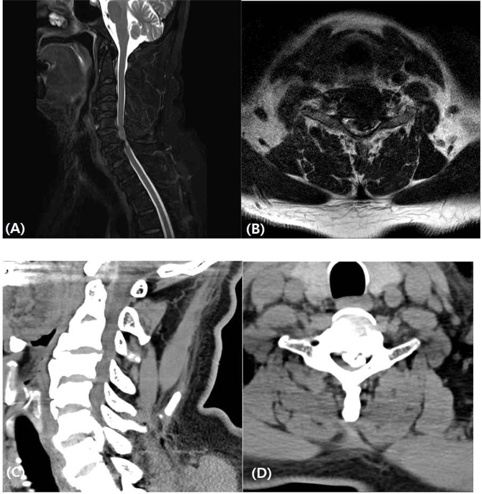
본 61세 남자 환자는 4개월 전부터 발생한 왼쪽 상하지 감각 이상과 왼쪽 하지 위약으로 본원 정형외과에 내원하였다. 영상 검사상 경추 6–7번에서의 후종인대골화증으로 인한 퇴행성 경부 척수증 진단 하에 IONM을 동반한 전방접근법(anterior approach) 경추 5–6–7번 전방경추 척추체제거술 및 유합술(anterior cervical decompression and fusion, ACDF, C6 corpectomy)을 계획하였다(Fig. 1).
Fig. 1.
Preoperative T2 weighted magnetic resonance imaging sagittal (A), axial (B) and computed tomography sagittal (C), axial (D) images of cervical spine.
Image showing focal ossification of the posterior longitudinal ligament at C6-7, markedly compressing spinal cord.
Download Original Figure
수술 하루 전, 운동유발전위 및 체성감각유발전위를 포함한 신경근전도검사를 양측 상하지에서 실시하였으며, 양하지 운동유발전위의 중추운동전도시간(central motor conduction time) 지연과 왼쪽 하지의 체성감각유발전위의 지연이 있었지만 유발은 되는 것을 확인하였다.
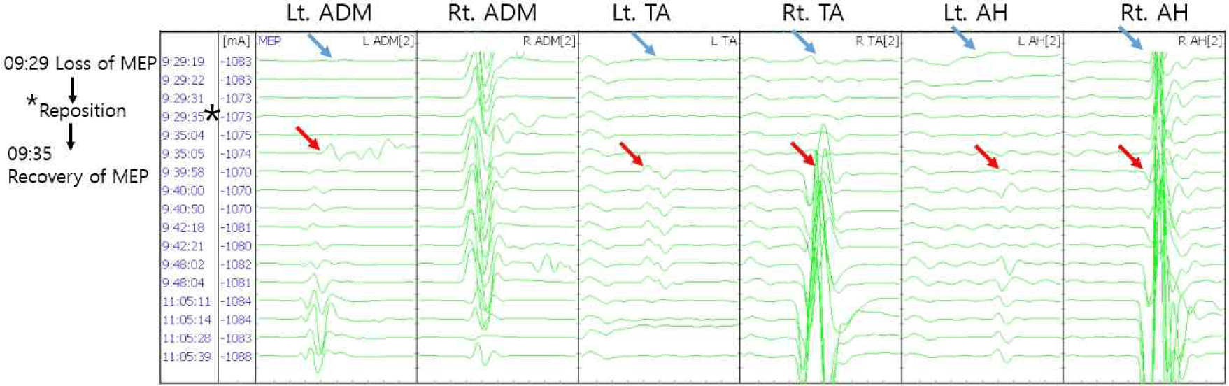
수술 당일, 누운(supine) 자세에서 전 정맥 마취(total intravenous anesthesia) 하에 마취는 시작하였고, 이후 수술이 시작하기 전, IONM 팀은 모니터링 장비를 연결(setup)하였다. 체성감각유발전위는 상지의 경우 양측 정중 신경(median nerve)에서 자극(stimulation)하여 C3’, C4’에서 측정(recording)하였으며, 하지의 경우 양측 경골 신경(tibial nerve)에서 자극하여 CZ’, FPZ’에서 측정하였다. 운동유발전위는 C3, C4에서 자극하여 양측 삼각근(deltoid), 이두근(biceps), 삼두근(triceps), 소지외전근(abductor digiti minimi), 전경골근(tibialis anterior), 무지외전근(abductor hallucis)에서 기저 운동유발전위(baseline MEP)를 측정하였다. 마취 시작 후 1시간 9분경 기저 운동유발전위는 모두 유발되었으나, 이후 환자는 수술을 위해 누운 자세에서 환자를 목을 신전(neck extension)시키고, 양쪽 어깨와 팔을 아래로 당긴 자세를 취하였다. 자세를 잡고 나서 10분 뒤 유발전위를 확인했을 때 대부분의 근육에서 운동유발전위 파형(wave form)이 유발되지 않았다(Figs. 2 and 3). IONM에 영향을 미칠 수 있는 모든 기술적 원인을 확인하였고, 수술 전 근전도 검사와, 자세잡기 전의 모니터링 과정에서 운동유발전위를 확인할 수 있었던 환자였기에, IONM 팀은 집도의에게 수술 자세로 인한 경부 척수 손상 가능성을 전달하였다. 수술의사는 즉시 수술 진행을 중단하고 환자의 목 자세를 조정하였고, 약 5분 후 모든 운동유발전위 파형이 회복되었으며 이를 확인 후 다시 수술을 진행하였다.
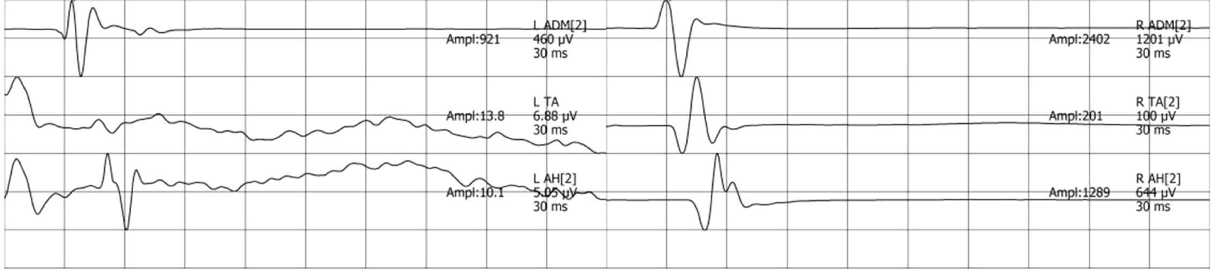
Fig. 2.
Baseline MEP of both ADM, TA, and AH muscle at the start of the surgery and prior to neck positioning.
MEP: motor evoked potentials; ADM: abductor digiti minimi; TA: tibialis anterior; AH: abductor halluces.
Download Original Figure
Fig. 3.
During the operation, intraoperative MEP signals were recorded.
After neck positioning, we observed a decline in the MEP amplitude of the left ADM, TA, and AH muscle. The surgeon adjusted the patient’s positioning (asterisk). The MEP of the most muscles showed low amplitude compared to baseline (blue arrow). The MEP of the left ADM, TA, and AH muscle showed signs of recovery, but the amplitude of the signals remained relatively low (red arrow). MEP: motor evoked potentials; Lt.: left; ADM: abductor digiti minimi; Rt.: right; TA: tibialis anterior; AH: abductor halluces.
Download Original Figure
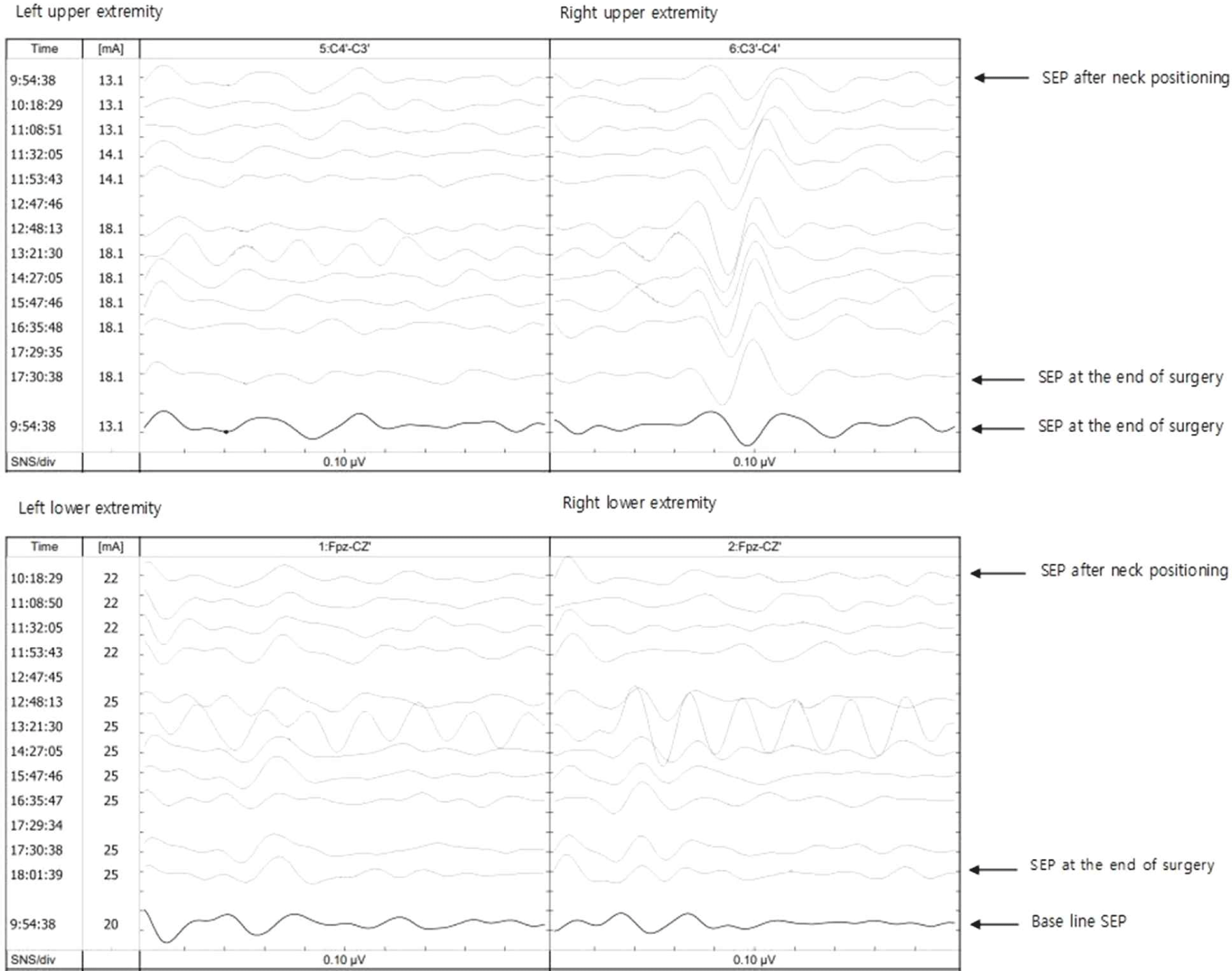
18시 20분에 수술은 종료되었으며, 이때 확인한 환자의 운동유발전위 신호는 모든 근육에서 유의미한 차이 없이 관찰되었으며, 좌상지를 제외한 체성감각유발전위의 진폭과 지연시간은 기저(baseline) 상태 비교하여 유의한 진폭 하락 또는 지연 없이 유지되었다. 좌상지에서는 진폭이 30% 감소하고 지연시간이 5% 지연되었다(Fig. 4). 수술 후, 모든 근육에서 5등급(normal)의 근력이 유지되었지만, 우측 상지의 근위부 영역에서 감각 이상이 발생했다. 감각 이상 증상은 퇴원 시 개선되었으며, 최종 추적 검사에서도 근력 저하가 발견되지 않았다.
Fig. 4.
During the operation, intraoperative SEP signals were recorded.
The amplitudes and latency of sensory-evoked potentials, except for the left upper limb, were maintained without significant amplitude reduction or latency delay compared to the baseline state (bold line). However, in the left upper limb, there was a 30% decrease in amplitude and a 5% delay in latency. SEP: somatosensory evoked potential.
Download Original Figure
고찰
이 증례 보고는 퇴행성 경부 척수증환자의 경추 수술에서 IONM의 사용이 잠재적인 신경 손상을 방지하는 데 중요하다는 점을 보여준다. 또한 수술 자체의 손상의 위험뿐 아니라, 수술을 준비하는 단계에서부터 부적절한 목 자세로 인한 신경 손상의 잠재적 위험을 증가시킬 수 있으며 이로 인해 합병증을 일으키고 수술 결과에 영향을 미칠 수 있다. 따라서 수술의 안전과 효과를 보장하기 위해 훈련된 모니터링 담당 의사 팀이 필요하다. 수술 전 근전도 검사와 수술 전 자세를 잡는 단계에서부터 IONM의 사용은 합병증의 위험을 최소화하고 환자 결과를 향상시키기 위한 척추 수술의 표준적인 방법으로 고려되어야 한다.
경부 척수증의 수술 중 IONM의 주요 이점은 영구적인 신경 손상을 예방 및 감소시키기 위해 수술 도중 보정 조치를 취할 수 있다는 것이다[5]. 이번 사례에서 강조된 바와 같이, 수술 전 목이나 팔의 위치 조정 중에도 신경 손상이 발생할 수 있으므로 목의 위치 조정을 포함하여 수술 전체에 걸쳐 IONM을 적용하는 것이 필수적이다. 수술 전 운동유발전위와 체성감각유발전위와 같은 검사는 환자의 기저 신경학적 상태를 평가하고 수술 도중 발생 가능한 신경 손상의 잠재적 위험 요인을 파악하는 데 매우 중요하다. 또한 수술 전 검사는 환자의 운동 및 감각 기능에 대한 유용한 정보를 제공하며 수술 접근 방식을 계획하는 데 도움이 된다[6]. 또한 일반적으로 시행되는 임상 검사에서는 발견되지 않을 수 있는 기존의 신경학적 결함을 확인하는 데 도움이 된다. 만약 모니터링이 수술실 안에서만 이루어진다면, 유발전위의 소실 혹은 지연이 환자의 기존 상태 때문인지 수술 중 발생한 사건 때문인지 구별하기 어렵다. 우리 케이스에서는 수술 전에 유발전위 검사를 통해 환자의 신경 상태를 평가하고, 이 데이터를 사용하여 수술실에서 확인된 비정상적인 데이터가 새롭게 발생한 문제인지를 판단했다. 따라서, 환자는 물론 수술의사의 자가 보호(self-protection)를 보장하기 위해 수술 전에 모니터링 팀과의 공동 작업(co-work)을 통해 환자를 평가하는 것이 중요하다.
또한, IONM의 이점은 수술 후에도 이어질 수 있다. 경추 수술을 받는 환자들은 종종 수술 이후에도 장기간의 치료와 재활이 필요하며, 이 과정에서 신경 모니터링은 회복 과정을 추적하고 추가적인 신경손상을 예방하는 데 중요한 역할을 할 수 있다[7]. 따라서, 수술 후 치료 계획 수립을 위해서 IONM을 시행하는 것이 중요하다.
결론
이 증례 보고는 경부 척수증 수술에서 IONM의 중요성을 강조하고, 수술 전 자세를 잡는 과정까지 모니터링을 해야 한다는 것을 보여준다. IONM의 사용은 신경 손상을 예방하고 수술 결과를 향상시킬 수 있다. 또한, 모든 경부 척수증 수술 환자에 대해 수술 전 유발전위 검사를 고려해야 한다.