
Introdcution
In spine surgery, motor evoked potentials (MEP) represent the most commonly utilized intraoperative neuromonitoring method. Among the various techniques, transcranial electrical stimulation motor evoked potentials (TES-MEP) is the most prevalently employed [1].
Anesthetics inducing muscle relaxation are frequently used in various surgical procedures that necessitate general anesthesia, primarily to prevent involuntary patient movement during the operation. However, for effective acquisition of TES-MEP in muscles, it is advisable to avoid anesthetics with muscle relaxant properties, such as halogenated gas anesthetics. Instead, total intravenous anesthesia (TIVA) is recommended to ensure stable MEPs in muscles. Nevertheless, instances of patient movement may still arise during TIVA. If the surgeon determines that maintaining the patient’s immobility during surgery takes precedence over neuromonitoring, they may opt to forgo intraoperative neurophysiological monitoring (IONM) and administer muscle relaxants at sufficient concentrations. Additionally, some institutions continuously use muscle relaxants at low concentrations while monitoring the train-of-four.
In surgical procedures, unexpected patient movements can risk nerve or blood vessel damage. Additionally, a sudden contraction of the masseter muscles can lead to the patient inadvertently biting the endotracheal tube, thereby impending proper aeration and adversely impacting oxygen saturation. Conversely, if the endotracheal tube quickly returns to its original shape after being bitten, there may be no impact on saturation. Therefore, detecting a biting event solely through oxygen saturation monitoring during surgery can be challenging. Such occurrences may further result in harm to the teeth or tongue. Patients may involuntarily move due to intense pain from unavoidable surgical stimuli or intentional electrical stimulations for MEP wave acquisition. TES, known to cause facial muscle contractions, poses a risk of oral injury. Therefore, utilization of a bite block is crucial for TES-MEP. It is noteworthy that the majority of IONM software systems incorporate alerts, prompting users to confirm the correct placement of the bite block placement prior to commencing TES.
Despite these preventive measures, tongue bite injuries can still occur. Here, we present two cases of tongue laceration subsequent to spinal surgery conducted under IONM.
Case 1
A 62-year-old male patient presented with hypoesthesia and paresthesia in both hands. He exhibited progressive bilateral hand and leg weakness, which led to gait disturbance. Despite these symptoms, his motor score, as assessed by the Medical Research Council (MRC) scale (using the 10 key muscles defined by the International Standards for Neurological Classification of Spinal Cord Injury), yielded a score of 100, indicating no definite preoperative weakness. Cervical MRI findings revealed cervical myelopathy associated with a herniated cervical disc and foraminal stenosis at C4/5 level. Consequently, surgical decompression was planned. The patient’s height and weight were 163.5 cm and 63.0 kg, resulting in a body mass index of 23.6. A posterior laminoplasty of C3 through 7 was planned with IONM.
The IONM encompassed transcranial electrical stimulation MEPs and free running electromyography (frEMG) of the bilateral deltoid, abductor pollicis brevis, tibialis anterior (TA), and abductor hallucis muscles. A single train of stimulation, consisting of 6 consecutive pulses (pulse width of 0.05 msec; interpulse interval of 3.0 msec), was applied at an intensity of 300–400 V using the interhemispheric montage of C3 and C4. The baseline waves were acquired using supramaximal stimulation at 300 V after confirming a train-of-four activity counted of four. The SEPs from the bilateral median nerves were recorded using a stimulation of 12–15 mA (pulse width of 0.3 msec; frequency of 4.7 Hz) at the C3-C4 montage. SEPs from the tibial nerves were obtained with a stimulation of 16–20 mA (pulse width of 0.3 msec; frequency of 4.7 Hz) using the Cz-Fz montage.
For general anesthesia, TIVA was employed. A smooth intubation was facilitated using a one-time intravenous dose of 50 mg of rocuronium, without subsequent administration of muscle relaxants. Following intubation in the supine position, a 1.5 cm diameter and 5 cm length bite block (Stick Bite Block, Soosung, Yangsan, Korea) was aligned with the endotracheal tube and fastened with a silicone tape. After intubation, the patient was maintained in a prone position throughout the surgical procedures.
During the foraminotomy of Lt. C6-7, an abrupt reduction exceeding 90% in the MEP amplitude of Lt. TA muscle was observed compared to the baseline (Fig. 1). This reduction persisted without subsequent recovery until the end of the monitoring. In an attempt to identify if waveforms could be acquired with a higher stimulation intensity, the intensity was incrementally increased to 400 V. Nonetheless, no MEP wave was recorded in the Lt. TA muscle. Throughout all surgical procedures, the SEPs remained stable without any signs of deterioration.

During surgery, crucial vital signs, such as oxygen saturation, blood pressure, respiration rate, and pulse rates were continuously monitored, and there was no significant deterioration in any of these essential parameters.
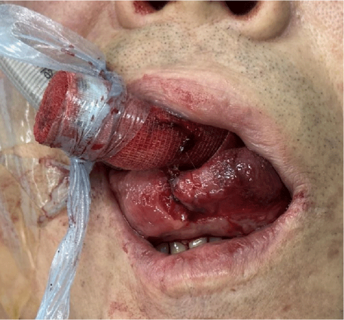
After completion of the surgical procedure, the patient was repositioned into a supine orientation. A subsequent oral examination revealed an intraoral hemorrhage stemming from a laceration located medially on the tongue, measuring approximately 2.5 cm in length and 1 cm in depth (Fig. 2). The lesion was immediately addressed and sutured by a dentist prior before the patient exited the surgical suite. No additional post-operative hemorrhagic complications were noted.
Two days post-surgery, the patient’s motor scores for Lt. shoulder abduction and elbow flexion were assessed to be grade 3. This finding is indicative of postoperative C5 palsy. Consequently, the patient was transferred to the Department of Rehabilitation Medicine for a targeted rehabilitation program.
Case 2
A 53-year-old man presented with a constellation of symptoms, including hypoesthesia, tingling sensations in both hands, weakness, and gait disturbance. He was diagnosed with cervical myelopathy at the C3-6 level, attributable to a herniated cervical disc at C4-5. This was further complicated by bilateral foraminal stenosis at C4-5 and on the right at C5-6. Upon evaluation, MRC motor score of 4 was determined for the key muscles corresponding to the Lt. C7, C8, and T1 nerve roots.
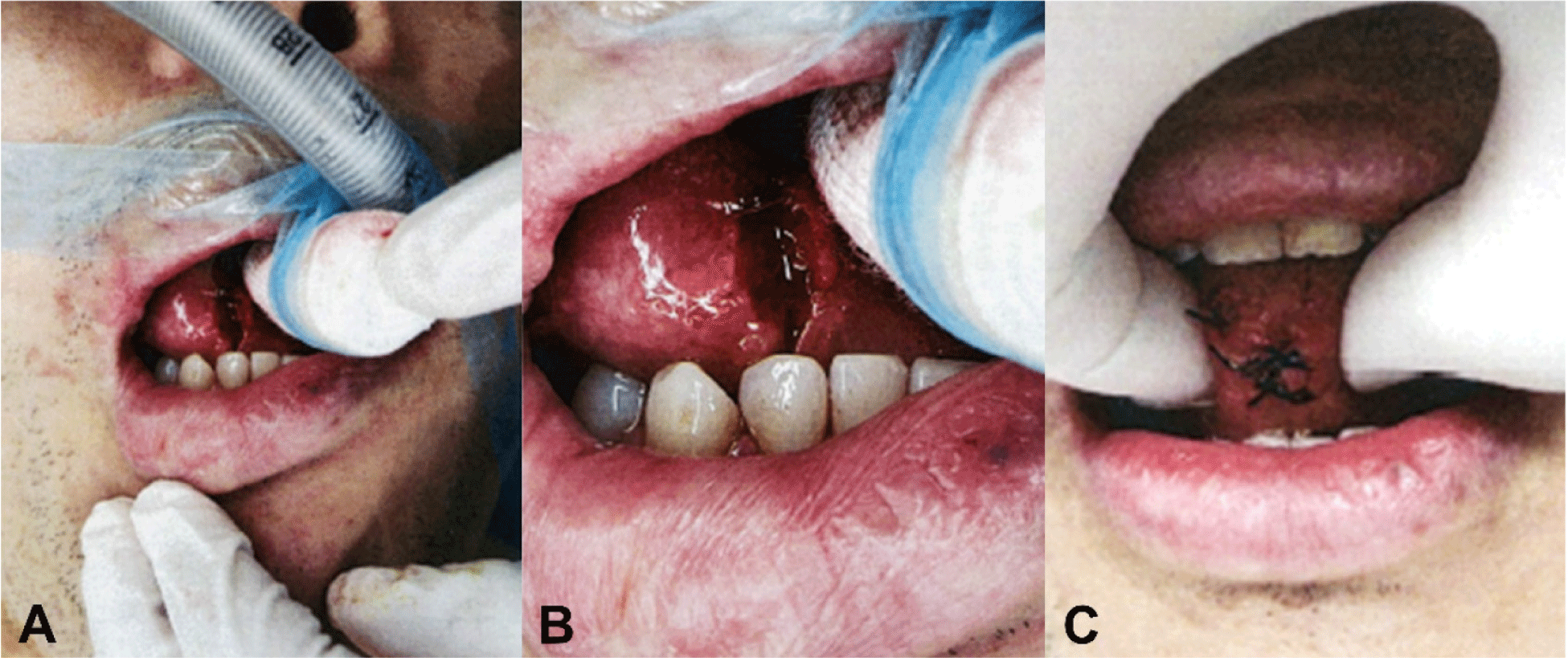
For general anesthesia, TIVA was employed. A smooth intubation was facilitated using a one-time intravenous dose of 50 mg of rocuronium, without subsequent administration of muscle relaxants. With the patient in a supine position after intubation, a 1.5 cm diameter and 5 cm length bite block (Stick Bite Block, Soosung) was aligned with the endotracheal tube and fastened with a silicone tape (Fig. 3-A). After intubation, the patient was maintained in a prone position throughout the surgical procedures. Posterior laminoplasty of C3 through 6 was performed under the guidance of IONM. MEPs, frEMG, and SEPs were continuously monitored throughout the entire surgical procedures, as described in Case 1. The intensity of TES for MEPs did not exceed 400 V. No significant deterioration was observed across all modalities.
During surgery, crucial vital signs, such as oxygen saturation, blood pressure, respiration rate, and pulse rates were continuously monitored, and there was no significant deterioration in any of these essential parameters.
Upon completion of the surgical procedures, the patient was repositioned into a supine orientation. Subsequent examination revealed a tongue laceration measuring approximately 1 cm (Fig. 3-B). The injury was promptly addressed and sutured by a dentist prior to the patient’s departure from the surgical suite (Fig. 3-C). No further hemorrhagic complications were observed. Postoperatively, the patient exhibited no deterioration in motor or sensory functions.
Discussion
Despite advancements in medical technology and the dedicated efforts of clinicians, instances of bite injuries are still sporadically reported. Historically, the incidence rate of bite injuries during surgeries using IONM has varied: 111/17,273 (0.63%), 29/15,000 (0.19%), 27/10,000 (0.27%), and 5/8,200 (0.06%), with our institute recording 2/7,000 (0.02%) [2,3]. Tongue laceration, followed by suturing, can lead to swelling and subsequent shortening of the tongue muscle. This may lead to restricted tongue movement, which is vital for propelling food to the posterior oral cavity and pharynx. Impaired tongue movement can hinder the ability to retain food within the oral cavity, thereby increasing the risk of aspiration. While tongue massage and exercises can aid in restoring function, it is imperative to prevent such injuries in the first place.
The precise cause of tongue laceration, whether attributed to TES or another surgical intervention, remains ambiguous in recent reports. Given the established association of TES with oral injuries, it is imperative for both IONM professionals and anesthesiologists to recognize and account for this risk. Kothbauer et al. mentioned that C3/4 stimulation is more likely to result in bite injury compared to C1/2 stimulation, due to its effect on the more intensive contraction of the temporalis muscles [4]. The C3/4 stimulation, with its deeper reach into the brain, has its merits for MEP monitoring of the lower limbs. As such, this technique is often used in spinal surgeries where MEP monitoring of the lower extremities is beneficial. Given the associated bite injury concerns, it is advisable for clinicians to exercise caution and be mindful of potential TES-related bite injuries. However, the double train stimulation technique is commonly employed in actual monitoring when there is a reduction in MEP, as it helps to ascertain the presence and extent of neural damage. Therefore, meticulous examination of the bite block is imperative for patients undergoing IONM.
Repeated TES can also present a risk of bite injuries. If TES is administered repeatedly without sufficient intervals, the effects of the preceding and subsequent stimulations can summate, exceeding the set stimulation value. This can significantly induce muscle contraction, and the masseter muscle is no exception.
Tongue bite injuries are more prevalent in the prone position because gravity can cause the tongue to protrude over the teeth, as presented in the aforementioned cases. Spinal surgeries with a posterior approach require patients to be in this position. In a previous study, 16 out of 17 bite injury events occurred during surgeries with a posterior approach [5]. In such cases, it is crucial for clinicians to carefully verify the placement of the bite block. This is especially significant for patients with a tongue that is disproportionately large or lengthy for their oral cavity, as there is an increased likelihood of the tongue positioning between the teeth [6].
In response, when an IONM specialist activates the MEP stimulation, most IONM software systems are designed to prompt an alert for the placement of a bite block before the actual TES deployment.
The bite block serves a critical role in preventing a range of bite injuries, encompassing those to the lips, oral cavity, and teeth. A notable case report even documents a mandibular fracture occurring when TES stimulation was administered without a bite block [7]. Bite injuries can result from inappropriately sized bite blocks or their displacement. Hence, it is essential for the bite block to be adequately large, ensuring the separation of teeth from the tongue, and securely fastened to minimize the risk of displacement. Clinicians have endeavored to enhance bite block designs to safeguard patients’ oral structures and accommodate endotracheal tubes.
Bite blocks can be categorized into two primary classifications: hard and soft. While the hard type effectively protects the endotracheal tube, it poses a higher risk of causing tooth injuries. To mitigate this risk, Yoshida et al. have proposed the use of a soft-type bite block made of spongy material [5]. A soft bite block made of gauze fashioned into a cylindrical shape is widely used, and ready-made products with this design are also available. In the instances of the current report, a piece of rolled gauze was positioned between the teeth. Given that a bite block with a diameter of 1.5 cm and a length of 5 cm was insufficient to prevent bite injury, plans are underway to use two rolled gauzes in the future. However, considering that the same size of bite block succeeded in preventing oral injury in some patients but failed in these two cases, it suggests the need to vary the type or size of the bite block depending on the patient. Oshita et al. previously designed a bite block using vinyl-silicone, by first making a mold of the patient’s teeth before the procedure [6]. This allows the endotracheal tube to pass through while also protecting the patient’s teeth. Nevertheless, this approach is time-consuming, costly, and not commonly used due to its inconvenience so far. There seems to be a need for the development of a more efficient, customized soft bite block.
In conclusion, a rare tongue injury was observed in these cases during TES-MEP monitoring, which can greatly affect a patient’s quality of life. Thus, designing an individualized bite block may be essential to prevent such incidents.