Case Report

전방 경추 추간판 제거술 및 유합술에서 수술중신경계감시의 유용성

이정윤1https://orcid.org/0000-0002-3544-1098, 백설희1,*https://orcid.org/0000-0002-3656-1833
Jungyeun Lee1https://orcid.org/0000-0002-3544-1098, Seol-Hee Baek1,*https://orcid.org/0000-0002-3656-1833
Author Information & Copyright
1고려대학교 안암병원 신경과학교실
1Department of Neurology, Korea University Anam Hospital, Korea University College of Medicine, Seoul, Korea
*Corresponding author: Seol-Hee Baek, Department of Neurology, Korea University Anam Hospital, Korea University College of Medicine, Seoul, Korea, Tel: +82-2-920-6285, Fax: +82-2-926-5347, E-mail: Virgo3318@gmail.com

© Copyright 2020 Korean Society of Intraoperative Neurophysiological Monitoring. This is an Open-Access article distributed under the terms of the Creative Commons Attribution Non-Commercial License (http://creativecommons.org/licenses/by-nc/4.0/) which permits unrestricted non-commercial use, distribution, and reproduction in any medium, provided the original work is properly cited.

Received: Feb 01, 2020 ; Revised: Feb 28, 2020 ; Accepted: Mar 09, 2020

Published Online: Mar 31, 2020

ABSTRACT

Anterior cervical discectomy and fusion (ACDF) surgery is the most common cervical spine surgery with generally favorable outcomes. However, the utility of intraoperative neurophysiological monitoring (INM) for ACDF surgery is still questionable. We report a case with the herniation of nucleus pulposus at C5, 6 levels who underwent ACDF surgery on INM. During surgery, the decreased amplitude of bilateral posterior tibial somatosensory evoked potential (SSEP) was detected. Then, SSEP was recovered after the administration of intravenous methylprednisolone. After surgery, this case had no neurological complications without paresthesia of the left hand. INM could be a useful tool in ACDF surgery.

Keywords: anterior cervical discectomy and fusion; evoked potentials, motor; evoked potentials, somatosensory; intraoperative neurophysiological monitoring; methylprednisolone

서론

수술중신경계감시(intraoperative neurophysiological monitoring)는 전기생리학적 검사들을 이용하여 수술 중 발생하는 신경손상을 조기에 확인하고 적절한 대처를 할 수 있게 하여, 수술 후 신경계 합병증을 줄이는데 도움을 줄 수 있는 검사이다[1]. 수술중신경계감시는 다양한 신경계 수술에서 적용할 수 있다. 척추수술(spine surgery) 분야에서는 척추측만증(scoliosis), 척수종양수술 등에서 수술중신경계감시를 시행하여 수술 후 합병증을 줄이는데 유용하게 활용되고 있다[2]. 전방 경추 추간판 제거술 및 유합술(anterior cervical discectomy and fusion)은 가장 널리 시행되는 경추 수술 중 하나이며, 약 0.4%에서 수술 후 신경계합병증이 발생한다고 보고되었다[3]. 전방 경추 추간판 제거술 및 유합술 시 수술중신경계감시의 유용성은 아직까지 명확하게 확립되지 않았다. 경추 수술에서 수술 중 신경손상을 확인하는데 수술중신경계감시가 도움이 된다는 연구 결과도 보고되었으나[4,5], 다른 연구에서는 전방 경추 추간판 제거술 및 유합술에서 수술중신경계감시를 시행한 군과 그렇지 않은 군에서 수술 후 신경계합병증의 빈도는 차이가 없다는 결과를 보고하기도 하였다[6].

저자들은 전방 경추 추간판 제거술 및 유합술에서 신경손상을 조기에 확인하고 대처하는데 수술중신경계감시가 유용하게 활용되었던 증례를 보고하고자 한다.

증례

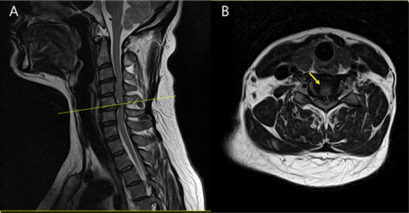
약 1년 전부터 경부 통증과 어깨 및 상지의 방사통이 있던 47세 여자 환자가 내원 2주 전부터 악화된 경부 통증을 주소로 내원하였다. 신경학적 검진에서 좌측 상지에서 Medical Research Council 등급 Ⅳ+로 관찰되었으며, 호프만징후 양성 소견이 확인되었다. 타원에서 시행한 경추 자기공명영상검사에서 경추 5/6위치의 추간판이 탈출되어 척수를 압박하고 있는 소견이 관찰되었다(Fig. 1). 상기 병변에 대해 경추 5/6번 추간판에 대해 전방 경추 추간판 제거술 및 유합술을 시행하고, 경추 4–6 위치에서 후궁성형술(laminoplasty)을 그리고 경추 3, 7번 위치에서 아전후궁절제술(subtotal laminectomy)을 시행하기로 계획하였다. 마취는 프로포폴(propofol) 3–8 μg/mL와 레미펜타닐(remifentanil) 2–8 ng/mL를 사용하여 완전정맥마취(total intravenous anesthesia)를 시행하였다. 기관 삽관을 위해 근이완제 로쿠로늄(rocuronium) 50 mg을 1회 정맥주사하였고, 이후 수술 과정에서는 사용하지 않았다. 수술중신경계감시는 Cascade® Elite(CADWELL Industries Inc., Kennewick, WA, USA) 장비를 이용하여 검사하였으며, 운동유발전위(motor evoked potential, MEP), train of four(TOF), 체성감각유발전위(somatosensory evoked potential, SSEP)와 자발근전도(spontaneous electromyography)를 이용하여 수술중신경계감시를 시행하였다. MEP는 두개경유전기자극(transcranial electrical stimulation) 방식으로 C3-4 위치에서 400 V의 5번 반복 펄스를 이용 자극하여 양측 새끼벌림근(abductor digiti quinti), 짧은엄지벌림근(abductor pollicis brevis), 정강앞근육(tibialis anterior), 무지외전근(abductor halluces)에서 기록하였다. TOF는 정중신경을 자극하여, 자극강도는 20 mA, 자극빈도는 2.73으로 자극하였다. SSEP는 양측 정중신경과 후경골신경을 자극하고, C3’-C4’, C4’-Fz에서 기록하였다. 자극 강도는 상지는 10 mV, 하지는 15 mV로 하며, 자극빈도 2.73 Hz로 자극하고 평균화 횟수는 150번으로 하였다. 자발근전도는 양측 위팔두갈래근(biceps brachii), 위팔세갈래근(triceps brachii), 노쪽손목굽힘근(flexor carpi radialis), 승모근(trapezius), 짧은엄지벌림근(abductor pollicis brevis), 정강앞근육(tibialis anterior), 무지외전근(abductor halluces)에서 기록하였다.

join-2-1-67-g1
Fig. 1. Cervical spine magnetic resonance imaging of the patient. (A) T2 weighted sagittal view and (B) T2 weighted axial view revealed disc herniation at C5/6 level (yellow dot-line and yellow allow).
Download Original Figure

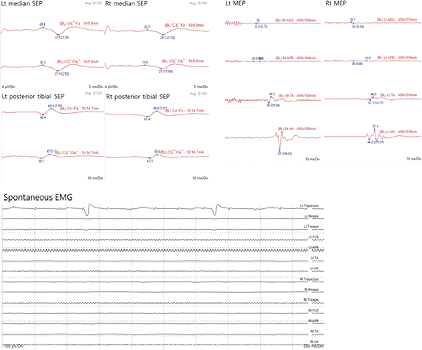
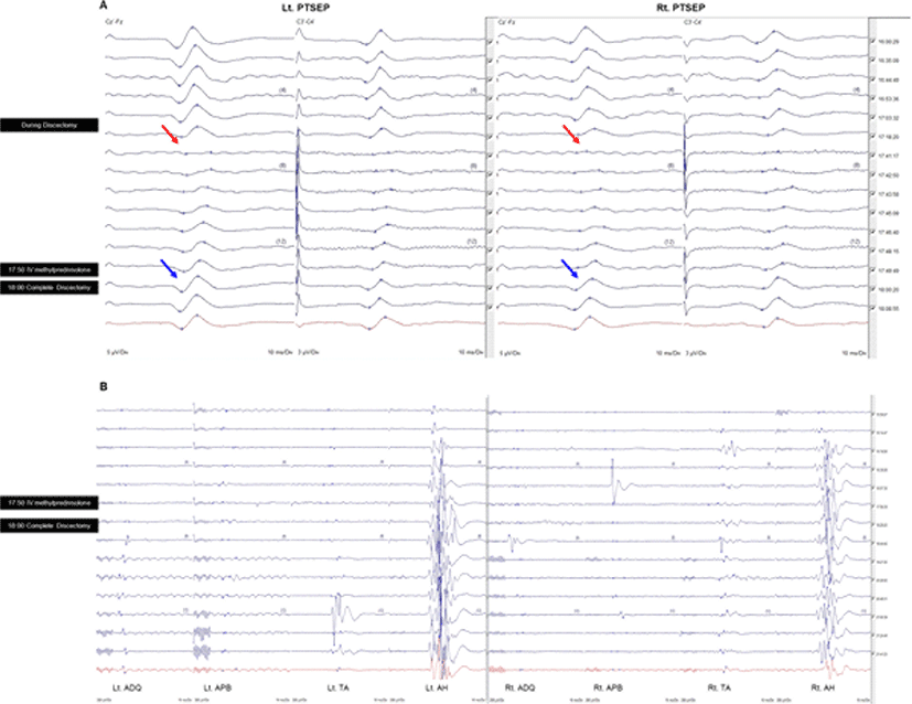
수술 시작 시 MEP 파형은 TOF 4/4인 상태에서 측정하였으며, 양쪽 무지외전근에서만 복합근활동전위(compound muscle action potential, CMAP)의 진폭이 작게 관찰되었고, 이외 부위에서는 MEP 파형이 형성되지 않았다(Fig. 2). 수술 시작 시 SSEP 파형에서는 특이소견은 관찰되지 않았다. 디스크를 제거하는 도중 양측 후경골신경 SSEP P37 파형의 진폭이 50% 이상 감소되는 소견이 관찰되어, 이에 대해 집도의에게 알렸다. 당시에 환자의 활력징후에는 특별한 변화가 없었으며, traction의 문제도 없음을 확인한 후, 즉시 메틸프레드니솔론 125 mg을 normal saline 100 mL에 섞어 급속 정주하였다. 투여 직후 SSEP의 진폭이 회복되는 소견이 관찰되었고, 메틸프레드니솔론 투여 10분 후 감압 완료되었을 시점에는 SSEP의 파형이 완전히 회복되는 소견이 확인되었다(Fig. 3-A). 수술 종료 시 MEP 파형은 여전히 양쪽 무지외전근에서만 관찰되었으나, CMAP의 진폭이 수술 시작 시에 비해 증가된 소견을 보였으며(Fig. 3-B), SSEP 파형은 완전히 회복된 상태로 종료하였다. 수술 후 환자의 신경학적 증상은 수술 전 소견과 비교하여 좌측 상지 근력이 호전된 소견을 보였으며, 수술 한달 후에는 좌측 손끝 저림 증상 외에는 다른 신경학적 증상은 없었다.

join-2-1-67-g2
Fig. 2. The waves of somatosensory evoked potential (SSEP), motor evoked potential (MEP), spontaneous electromyography at the baseline. At the beginning of surgery, SSEPs and spontaneous electromyography were obtained stably. However, MEPs were not recorded except in both abductor halluces muscles. ADQ: abductor digiti quinti; APB: abductor pollicis brevis; TA: tibialis anterior; AH: abductor halluces; FCR: flexor carpi radialis.
Download Original Figure
join-2-1-67-g3
Fig. 3. (A) Significant changes in the amplitude of somatosensory evoked potentials (SSEPs). During a discectomy, the amplitudes of the SSEPs were significantly reduced (red arrow). The amplitude of SSEPs was recovered after the administration of intravenous methylprednisolone (blue arrow). (B) The amplitude of motor evoked potentials was slightly increased after the administration of intravenous methylprednisolone. ADQ: abductor digiti quinti; APB: abductor pollicis brevis; TA: tibialis anterior; AH: abductor halluces.
Download Original Figure

고찰

본 증례는 전방 경추 추간판 제거술 및 유합술 중 SSEP의 이상소견을 확인하고, 메틸프레드니솔론 정맥주사 후 다시 파형이 정상으로 회복되었으며, 수술 후 신경학적 소견의 악화는 관찰되지 않았다. 본 증례는 MEP, SSEP 및 자발근전도를 이용하여 수술중신경계감시를 계획하였으나, 수술 초기부터 MEP가 잘 형성되지 않았던 증례로, 수술 중 신경손상에 대한 감시를 위해 MEP보다도 SSEP에 대한 감시가 중요하였다.

전방 경추 추간판 제거술 및 유합술은 경추 신경뿌리병증 및 경추 척수병증의 치료를 위해 흔히 시행하는 수술법이다. 전방 경추 추간판 제거술 및 유합술 이후 신경계합병증은 일시적인 저림이나 이상감각부터 비가역적인 마비(paralysis)까지 다양하게 나타날 수 있다[3]. 미국에서 시행한 연구 결과, 전방 경추 추간판 제거술 및 유합술에서 수술중신경계감시는 신경계합병증을 낮추는데 유용하다(Odd ratio 0.6, 95% CI 0.47–0.76, p < 0.001)고 보고되었다[5]. 하지만, 다른 연구에서는 수술중신경계감시를 시행한 그룹과 그렇지 않은 그룹에서 신경계합병증의 발병율은 통계적으로 유의한 차이를 보이지 않았으며(0.17% vs. 0.22%, p = 0.41), 병원비는 유의하게 증가하는 것으로 나타났다[6]. 전방 경추 추간판 제거술 및 유합술에서 수술중신경계감시의 유용성에 대해서는 더 많은 증례를 바탕으로 연구가 필요할 것으로 생각된다.

척추 수술에서 MEP와 SSEP는 기본적으로 시행하는 검사이며, 일반적으로 MEP가 SSEP보다 더 민감한 검사로 알려져 있다. 척추수술에서 SSEP의 민감도는 약 22%–100%이고, 특이도는 100%로 추정되며, MEP의 민감도는 78%–100%, 특이도는 83.2%–100%로 추정된다[4]. 전방 경추 추간판 제거술 및 유합술 200 증례를 분석한 연구 결과에서는 multi-channel MEP 검사는 SSEP보다 민감도, 특이도, 양성예측도 및 음성예측도가 모두 높은 소견을 보였다[7]. 이들 연구결과를 볼 때, MEP가 SSEP보다 척추수술에서 신경손상을 예측하는데 더 유용한 검사로 생각된다. 하지만 일부에서는 MEP의 변화 없이 SSEP만 변화를 보인 증례도 보고되었으며[8], 본 증례와 같이 초기부터 MEP가 형성되지 않는 경우에는 SSEP에 대한 감시가 중요하다.

급성 척수 손상의 병태 생리를 고찰하여 보면, 손상의 메커니즘은 세포막 기능 및 세포막의 lipase의 활성화와 관련이 있다. 이로 인해 이차적으로 염증 반응 및 부종, 허혈이 증가한다[9]. 메틸프레드니솔론과 같은 스테로이드가 신경 손상의 회복에 미치는 영향에 대한 기전에 대해서 아직까지 전부 밝혀진 것은 아니지만, 신경 조직의 전해질 균형을 유지해주고, 막의 안정화를 돕고, 세포 부종과 관련하여 수분과 이온의 균형을 조절하는 등 이로운 역할을 할 수 있다[10]. SSEP의 유의한 변화를 유발할 수 있는 소견으로는 일차적으로 직접적인 신경손상뿐만 아니라, 혈압 강하나 주위 혈관이 당겨지거나 눌리면서 발생하는 허혈성 원인을 고려해 볼 수 있다. 이런 경우, 파파베린 같은 혈관 이완제를 투여해 볼 수 있다[11,12]. 하지만 본 증례에서는 SSEP 변화 당시, 활력징후는 안정적이었으며, 신경이나 혈관의 traction도 체크해 보았으나 특별한 이상 없어 이차적인 원인보다는 일차적인 직접 신경 손상 가능성을 염두하여 메틸프레드니솔론을 투여하였고, 이후 SSEP가 회복되는 소견이 관찰되었다.

스테로이드가 아직까지 척수 손상 환자에서 스테로이드의 투여에 대한 근거는 명확하지 않다. 하지만 급성 척수 손상과 경부 척수 질환의 수술적 치료에서 수술 중과 전후로 스테로이드 투여 후 결과가 좋게 나타났다고 보고되고 있다[1316]. 한 연구에서는 전방 경추 수술 시행 환자들을 덱사메타손 혹은 생리식염수로 무작위 배정하였고, 수술 절개 직전과 수술 8–16시간 이후에 투여하였고, 덱사메타손 투여군에서 삼킴곤란 증상이 의미 있게 호전된 것으로 나타났다[13]. 수술중신경계감시 하에 척수 수술 도중 MEP 파형의 진폭 감소 소견이 확인된 직후 고용량 메틸프레드니솔론을 투여하였고, 이후 MEP가 회복되는 증례도 보고되었다[14]. 급성 척수 손상에 대한 메타분석에서는 손상 8시간 이내에 메틸프레드니솔론 투여가 효과가 있다고 보고되기도 하였다[16]. 본 증례도 스테로이드가 신경손상의 회복에 도움이 될 수 있음을 보여주는 증례로 생각된다.

결론적으로, 전방 경추 추간판 제거술 및 유합술에서 수술중신경계감시는 수술 중 신경계 손상을 실시간으로 확인하면서 필요시 적절한 대응을 할 수 있다는 점에서 유용성이 더 클 것으로 생각된다. 향후 많은 증례들에 대한 추가적인 연구가 필요할 것으로 생각된다.

Ethical approval

This article does not require IRB/IACUC approval because there are no human and animal participants.

Conflicts of interest

No potential conflict of interest relevant to this article was reported.

참고문헌

1.

Fehlings MG, Brodke DS, Norvell DC, Dettori JR. The evidence for intraoperative neurophysiological monitoring in spine surgery: does it make a difference? Spine (Phila Pa 1976). 2010;35(9 Suppl):S37-46.

2.

Lall RR, Lall RR, Hauptman JS, Munoz C, Cybulski GR, Koski T, et al. Intraoperative neurophysiological monitoring in spine surgery: indications, efficacy, and role of the preoperative checklist. Neurosurg Focus. 2012;33(5):E10.

3.

Kashkoush A, Mehta A, Agarwal N, Nwachuku EL, Fields DP, Alan N, et al. Perioperative neurological complications following anterior cervical discectomy and fusion: clinical impact on 317,789 patients from the national inpatient sample. World Neurosurg. 2019; 128:e107-15.

4.

Di Martino A, Papalia R, Caldaria A, Torre G, Denaro L, Denaro V. Should evoked potential monitoring be used in degenerative cervical spine surgery? A systematic review. J Orthop Traumatol. 2019;20(1):19.

5.

Ney JP, Van der Goes DN, Nuwer MR. Does intraoperative neurophysiologic monitoring matter in noncomplex spine surgeries? Neurology. 2015; 85(24):2151-8.

6.

Badhiwala JH, Nassiri F, Witiw CD, Mansouri A, Almenawer SA, da Costa L, et al. Investigating the utility of intraoperative neurophysiological monitoring for anterior cervical discectomy and fusion: analysis of over 140,000 cases from the National (Nationwide) Inpatient Sample data set. J Neurosurg Spine. 2019;31(1):76-86.

7.

Kim DG, Jo SR, Park YS, Hyun SJ, Kim KJ, Jahng TA, et al. Multi-channel motor evoked potential monitoring during anterior cervical discectomy and fusion. Clin Neurophysiol Pract. 2017;2:48-53.

8.

Ahn JH, Park JJ, Oh DA, Yoon BN. A spinal cord tumor removal case with somatosensory evoked potential change more severe than motor evoked potential change. J Intraoper Neurophysiol. 2019; 1(2):44-9.

9.

Matsumoto T, Tetsuya T, Mamoru K, Munehito Y, Muneharu A, Hiroshi Y. Early complications of high-dose methylprednisolone sodium succinate treatment in the follow-up of acute cervical spinal cord injury. Spine (Phila Pa 1976). 2001; 26(4): 426-30.

10.

Young W, Flamm ES. Effect of high-dose corticosteroid therapy on blood flow, evoked potentials, and extracellular calcium in experimental spinal injury. J Neurosurg. 1982;57(5):667-73.

11.

Robertazzi RR, Cunningham JN. Intraoperative adjuncts of spinal cord protection. Semin Thorac Cardiovasc Surg. 1998;10(1):29-34.

12.

Rivlin AS, Tator CH. Effect of vasodilators and myelotomy on recovery after acute spinal cord injury in rats. J Neurosurg. 1979;50(3):349-52.

13.

Cui S, Scott D, John CF, Sanford EE. The effects of perioperative corticosteroids on dysphagia following surgical procedures involving the anterior cervical spine: a prospective, randomized, controlled, double-blinded clinical trial. J Bone Jt Surg. 2019;101(22): 2007-14.

14.

Jeon DG, Park YG, Cho YE, Kim D, Park J. Effects of intravenous methylprednisolone on intraoperative neurophysiological monitoring during spinal cord tumor surgery: a case report. J Intraoper Neurophysiol. 2019;1(2):50-4.

15.

Hugenholtz H, Cass DE, Dvorak MF, Fewer DH, Fox RJ, Izukawa DMS, et al. High-dose methylprednisolone for acute closed spinal cord injury--only a treatment option. Can J Neurol Sci. 2002;29(3):227-35.

16.

Bracken MB. Steroids for acute spinal cord injury. Cochrane Database Syst Rev. 2002;3:CD001046.