Case Report

후교통동맥 결찰술 중 수술중 신경계 감시에서 탐지되지 않은 시상핵 허혈성 손상

김진아1https://orcid.org/0000-0001-9378-346X, 김성민1,*https://orcid.org/0000-0003-2425-6388
Jin-Ah Kim1https://orcid.org/0000-0001-9378-346X, Sung-Min Kim1,*https://orcid.org/0000-0003-2425-6388
Author Information & Copyright
1서울대학교병원 신경과
1Department of Neurology, Seoul National University Hospital, Seoul, Korea
*Corresponding author: Sung-Min Kim, Department of Neurology, Seoul National University Hospital, 101 Daehak-ro, Jongro-gu, Seoul 03080, Korea, Tel: +82-2-2072-7312, E-mail: Sueh916@gmail.com

© Copyright 2020 Korean Society of Intraoperative Neurophysiological Monitoring. This is an Open-Access article distributed under the terms of the Creative Commons Attribution Non-Commercial License (http://creativecommons.org/licenses/by-nc/4.0/) which permits unrestricted non-commercial use, distribution, and reproduction in any medium, provided the original work is properly cited.

Received: Aug 20, 2020 ; Revised: Aug 31, 2020 ; Accepted: Sep 04, 2020

Published Online: Sep 30, 2020

ABSTRACT

Intraoperative neurophysiological monitoring during aneurysm surgery can reduce the risk of ischemic insults and can improve neurological prognoses by detecting abnormal findings in real time when cerebral circulation is at risk. In aneurysm clipping surgery, ischemic insults are most likely to occur at the clipping step. Somatosensory evoked potentials (SSEPs) are known to be the most sensitive method for detecting ischemic insults. However, there are several cases in which SSEP findings and the postoperative prognosis are inconsistent. we report a patient who showed postoperative neurologic deficits after posterior communicating artery (P-com) aneurysm clipping surgery, even though the amplitude of the SSEP had fully recovered shortly after the point at which it had decreased by 15%. The patient’s brain MRI revealed an anterior thalamic infarction, and transfemoral cerebral angiography showed an anterior thalamoperforating artery occlusion. This case shows that if a thalamic infarction occurs in the anterior region (not in the posterolateral region, which is involved in the somatosensory pathway), then it may not be identified by SSEP.

Keywords: aneurysm clipping; anterior thalamic infarction; evoked potentials, somatosensory; intraoperative neurophysiological monitoring

서론

뇌동맥류의 수술적 결찰술은 동맥류의 파열을 사전에 예방하여 치명적인 합병증으로부터 환자를 보호하는 것이 목적이다. 개두술을 통해 뇌동맥류를 노출하고, 미세 클립을 뇌동맥류 경부에 거치시켜 혈류를 차단하는 수술이며, 뇌동맥류의 파열위험이 높은 경우에는 근위부에 일시결찰(temporary clipping)을 한 후에 최종 결찰을 시행하게 된다[1]. 뇌동맥류 수술 과정 중에는 다양한 합병증이 발생할 수 있지만, 일시결찰 중에서 특히 뇌허혈 손상이 일어날 가능성이 높아 수술중신경계감시기법의 역할이 크다[2]. 체성감각유발전위(somatosensory evoked potential, SSEP)는 수술중신경계감시에서 가장 많이 사용되는 방법 중 하나로, 뇌혈관 수술에서 역시 그 활용도가 높다. 뇌동맥류 수술 중 결찰을 하는 동안 허혈성 변화를 실시간으로 감시하여 집도의로 클립 적용 시간을 조절하도록 함으로써 신경학적 예후를 개선시키는데 기여할 수 있다. 하지만 체성감각유발전위 소견과 실제 수술 후 예후와 일치하지 않는 경우들이 있는데[3], 이번 증례에서는 후교통동맥 동맥류 결찰술 중 체성감각유발전위의 진폭이 15%까지 감소했다가 비교적 빠른 시간 내에 호전되었음에도 불구하고, 신경학적 후유증이 남았던 사례를 보고하고자 한다.

증례

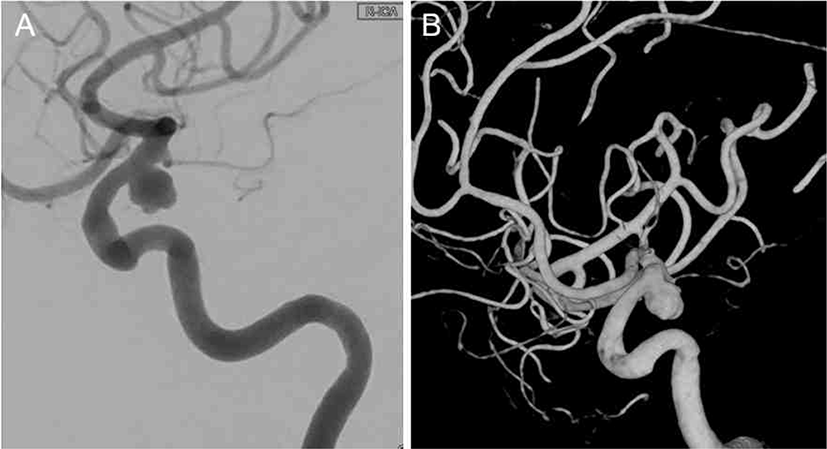
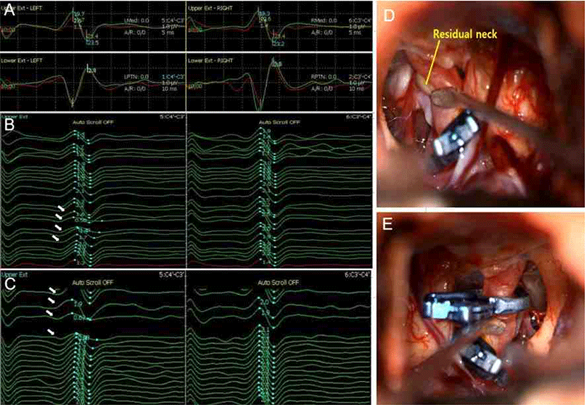
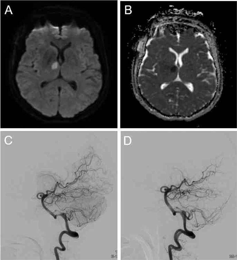
62세 여자 환자가 건강검진으로 시행한 뇌 MRI에서 우측 뒤교통동맥에 4 × 5의 뇌동맥류(Fig. 1)가 확인이 되어 결찰술을 시행하였다. 수술 전 신경학적 검사에서 이상 소견은 없었다. 마취는 프로포폴(propofol)과 레미펜타닐(remifentanil)을 사용하여 완전정맥마취(total intravenous anesthesia, TIVA)를 시행하였고, 수술중신경계감시기법으로는 SSEP, 운동유발전위(motor evoked potential, MEP), 자발근전도(spontaneous electromyography)를 사용하였다. SSEP는 정중신경(median nerve)과 정강신경(tibial nerve)에서 상지는 18 mA, 하지는 30 mA의 세기로 2.8 Hz의 빈도로 자극하고, C3-C4’, C4’-C3’, CZ’-Fpz에서 기록하였다. MEP는 상지에서는 양측 삼각근(deltoid), 양측 엄지두덩근육(thenar muscle), 하지에서는 양측 앞정강근(tibialis anterior), 양측 엄지발가락벌림근(abductor hallucis)에서 측정하였고, 자극은 두개경유전기자극(transcranial electrical stimulation) 방식으로 C3-C4 위치에서 350V의 세기로 6회 반복펄스로 자극하였다. 자발근전도는 양측 삼각근, 양측 엄지두덩근육, 양측 앞정강근, 양측 엄지발가락벌림근에서 측정하였다. 경막을 열고 10분 정도 지나 동맥류를 노출시켰고, 근위부의 모동맥을 확보하였다. 동맥류의 크기가 크고 조기파열 위험이 높아 모동맥의 일시결찰을 시행하였다. 우측 내경동맥(internal carotid artery)에 일시결찰을 하고, 우측 후교통동맥의 관통동맥(perforating artery)들을 박리하여 클립 날(blade)이 들어갈 공간을 확보하였다. 일시결찰 직후에 좌측 상, 하지 SSEP의 진폭(amplitude)이 15%까지 감소하였다. 뇌동맥류 경부에 영구결찰(permanent clipping)을 시행하고 일시결찰을 풀자 2분 후 SSEP가 기저상태로 회복되었다. 후교통동맥의 기시부와 전맥락막동맥(anterior choroidal artery)의 기시부가 손상되지 않았음을 확인하였으며, 관통동맥도 잘 보존되어 있었다(Fig. 2). 하지만 뇌동맥류 경부 앞쪽에 잔존낭(residual sac)이 보여, SSEP가 모두 회복되고 3분 후에 다시 우측 내경동맥에 일시결찰을 시행하고, 30초 후에 잔존낭에 대한 영구결찰이 시행되었다. 영구결찰 30초 후 일시결찰을 풀었고, 총 일시결찰 지속시간은 1분 35초로 확인되었다. 두 번째 일시결찰 시에도 결찰 약 1분 후 좌측 상, 하지 SSEP가 30%까지 감소하였으나 일시결찰 제거 후엔 모두 회복되었고, 수술 종료 시에도 SSEP의 변화 없이 마무리가 되었다(Fig. 2). MEP와 자발근전도에서는 유의미한 이상소견은 확인되지 않았다. 수술 5시간 후 환자 평가 시 시간, 장소, 사람에 대한 지남력이 떨어지고 밖에 사람이 있다고 반복적으로 이야기하는 섬망 증상을 보이기 시작하였다. 이 때 사지의 위약감이나 감각변화는 관찰되지 않았다. 수술 후 합병증을 평가하기 위해 뇌 확산강조영상(diffusion-weighted imaging, DWI)을 시행하였고, 앞시상핵에 국소적인 확산제한(diffusion restriction) 소견이 확인되었고, 확산계수(apparent diffusion coefficient, ADC)는 감소한 소견을 확인하였다. 이어 시행한 뇌혈관조영술(transfemoral cerebral angiography, TFCA)에서 수술 전에 개통이 잘 확인되었던 전시상관통동맥(anterior thalamoperforating artery)이 보이지 않는 소견이 확인되었다(Fig. 3).

join-2-2-114-g1
Fig. 1. A pre-operative conventional angiography image of the patient. (A) The patient had a 4 × 5 cm-sized unruptured aneurysm in right posterior communicating artery. (B) 3D reconstruction rotational angiography of the patient.
Download Original Figure
join-2-2-114-g2
Fig. 2. Intraoperative neurophysiological monitoring during aneurysmal clipping surgery in our case and operative field findings. (A) The red wave is baseline of the median nerve somatosensory evoked potential (MNSSEP) and green wave is the MNSEP at the end of surgery. (B) As soon as the temporary clipping was applied, the amplitude of the MNSEP decreased by 15%, and when the clip was released, it recovered to its baseline (white arrow). (C) The residual aneurysmal neck was observed, so additional clipping was performed. The amplitude of the MNSEP decreased to 30% again when the second temporary clipping was applied. And it recovered again when the temporary clipping was released after second permanent clipping was performed (white arrow). (D) The operative field finding after the first permanent clipping. Residual aneurysmal neck was observed (yellow mark). (E) The operative field finding after the second permanent clipping.
Download Original Figure
join-2-2-114-g3
Fig. 3. Diffusion-weighted MRI on the 5 hours after surgery. (A) About 1.5 cm-sized diffusion restricted lesion on anterior thalamus. (B) Apparent diffusion coefficient (ADC) maps showed dark intensity. (C) Transfemoral cerebral angiography (TFCA) performed before the surgery showed patent anterior thalamoperforating artery. (D) TFCA performed after surgery showed invisible anterior thalamoperforating artery.
Download Original Figure

고찰

본 증례는 뇌동맥 결찰 수술 중 일시결찰 단계에서 SSEP의 진폭이 15%까지 떨어졌다가 바로 회복되었던 증례로, 수술 후 신경학적 결손이 없을 것으로 예상하였으나, 허혈성 뇌손상이 발생하였던 사례이다. 비파열 뇌동맥류 결찰술은 동맥류의 파열을 사전에 예방하여 환자의 안전을 도모하는 목적으로 시행되며, 수술 전에는 대개 무증상인 경우가 많기 때문에 수술 후 합병증 예방에 더욱 많은 노력을 기울이게 된다. 따라서 수술중신경계감시의 역할이 특히 중요하다. 뇌동맥류 수술 중에 합병증이 발생할 수 있는 상황은 수술 중 과도한 견인으로 인한 뇌표면의 미세순환장애, 뇌동맥류 주위의 관통동맥 폐색, 일시결찰 중의 뇌허혈, 모동맥 협착으로 인한 뇌허혈 등이 있는데, 이 중 대개 결찰 단계에서 뇌손상 발생 가능성이 높다[2]. 수술중신경계감시 중 이상 소견이 발생하였을 때 실시간으로 집도의에게 바로 피드백을 주어 영구적인 신경 손상을 예방할 수 있다.

그 중 SSEP는 여러 수술중신경계감시 기법 중 민감도가 가장 높은 검사로 알려져 있는데, 한 후향적 연구에서는 뇌동맥류 수술에서 이상이 감지된 3.83%의 증례 중 88.9%가 SSEP에서 처음 확인되었다. 이 때 가역적인 변화(reversible change)에서는 약 90%에서 신경학적 후유증 없이 회복된 것으로 보고되었다[4]. 이번 증례에서는 일시결찰을 두 번 시행하였고, 두 번 모두 SSEP의 진폭이 50% 이상 감소하여 허혈 상태를 잘 반영하였던 것으로 판단된다. 하지만 두 번 모두 2분 이내로 결찰 시간이 짧았고, 일시결찰을 제거한 후 곧바로 회복되었기 때문에 허혈로 인한 신경 손상 합병증 가능성이 높지 않을 것으로 판단하였다. 하지만 본 증례의 환자는 앞쪽시상핵 급성 뇌경색이 확인되었고, 이는 시상핵 내에서도 SSEP가 담당하는 체성감각신경 주행경로에서 벗어난 허혈성 손상은 신경계 감시를 벗어날 수 있다는 것을 보여주었다.

SSEP는 체성감각 중 고유수용성 감각(proprioception), 진동 감각(vibration sense)를 담당하는 뒤기둥(dorsal column)의 경로를 평가한다. 말초신경에서 자극을 받게 되면 연수(medulla oblongata)에서 이차 신경세포와 시냅스를 형성하고, 교차 후 반대쪽 안쪽섬유띠(medial lemniscus)를 거쳐 시상의 뒤가쪽핵(posterolateral)에서 삼차 신경세포와 시냅스를 형성하고, 시상피질투사(thalamocortical projection)를 통해 일차감각피질(primary sensory cortex)에 도달한다[2]. SSEP는 감각신경계를 평가하지만, 운동신경계와 감각신경계의 위치가 근접하여 운동신경계의 변화가 감지될 만큼 민감도가 높다[5]. 하지만 이번 증례에서 앞쪽 시상핵의 허혈성 손상은 SSEP에 반영되지 않는 것으로 보아, 시상핵 단계에서 앞쪽 시상핵의 선택적인 손상은 뒤가쪽핵의 체성감각경로에는 반영되지 못함을 알 수 있다.

본 증례의 환자는 수술 후에 운동 기능과 감각기능의 이상 없이 섬망 증상과 지남력 감소의 혼동(confusion)을 보여 뇌 MRI를 시행하였다. 고령의 환자에서 간혹 수술 후에 일시적으로 섬망 증상을 보일 수는 있지만, 본 증례의 환자처럼 후순환혈관 수술을 한 경우에는 수술 후 시상핵의 손상 가능성을 염두에 두고 주의깊게 보아야 한다. 시상핵 앞쪽 혹은 안쪽에 손상이 생기면 의지 상실, 무감동, 지남력 장애가 발생할 수 있고[6], 앞쪽 시상핵은 후교통동맥에서 분지한 결절시상동맥 혹은 극시상동맥(tuberothalamic or polar artery)의 지배를 받기 때문에 후교통동맥의 동맥류 수술 시 손상을 받을 수 있다. 극시상동맥은 후교통동맥의 관통동맥으로 관통동맥 분지가 클립에 같이 물리거나, 수술 중 직접적인 손상, 허혈 손상 모두 가능하다[7]. 한 연구에 따르면 관통동맥 손상은 대개는 영구결찰 과정에서 발생하지만, 시상관통동맥(anterior thalamoperforating artery)에서는 영구결찰에서 50%, 일시결찰 과정에서 50% 정도 발생하는 것으로 알려져 있다[8]. 본 증례의 경우는 수술 중 어떤 단계에서의 손상인지 판단하기 어렵우나, 전시상관통동맥 영역이란 점을 고려할 때 일시결찰 중에 전시상핵 경색이 발생하였을 가능성이 높을 것으로 판단된다. 본 증례는, 후교통동맥 동맥류 결찰술에서 시상핵 손상이 생겼을 때 뒤가쪽시상핵을 벗어난 영역에서는 수술중신경계 감시에서 확인되지 않을 수 있다는 것을 보여준다.

Conflicts of interest

No potential conflict of interest relevant to this article was reported.

Ethical approval

This article does not require IRB/IACUC approval because there are no human and animal participants.

References

1.

Kim YB. Surgical process of cerebral aneurysm clipping. J intraoper Neurophysiol. 2020;2(1):27-32.

2.

Ghaly RF, Dujovny M, Charbel F, Slavin K, Ausman JI. Monitoring the intracranial aneurysm patient. Neurol Res. 1994;16(1):12-7.

3.

Thomas B, Guo D. The diagnostic accuracy of evoked potential monitoring techniques during intracranial aneurysm surgery for predicting postoperative ischemic damage: a systematic review and meta-analysis. World Neurosurg. 2017;103:829-40.

4.

Thirumala PD, Udesh R, Muralidharan A, Thiagarajan K, Crammond DJ, Cahng YF, et al. Diagnostic value of somatosensory-evoked potential monitoring during cerebral aneurysm clipping: a systematic review. World Neurosurg. 2016;89:672-80.

5.

Koo YS, Kim DY. Basic principles and practices of evoked potential for intraoperative neurophysiological monitoring: motor evoked potential and somatosensory evoked potential. J intraoper Neurophysiol. 2019;1(1):14-24.

6.

Rikhye RV, Wimmer RD, Halassa MM. Toward an integrative theory of thalamic function. Annu Rev Neurosci. 2018;8(41):163-83.

7.

Cha JH, Park IS, Yi J, Kim KJ, Hwang SH, Han JW. Ischemic complication after surgery for posterior communicating artery aneurysm. J Korean Neurosurg Soc. 2003;33:137-41.

8.

Sasaki T, Kodama N, Matsumoto M, Suzuki K, Konno Y, Sakuma J, et al. Blood flow disturbance in perforating arteries attributable to aneurysm surgery. J Neurosurg. 2007;107(1):60-7.