서론
경추 압박성척수증(cervical compressive myelopathy)은 척수의 기능장애를 일으키는 대표적인 원인으로 추간판 탈출, 척추증(spondylosis), 후종인대골화증(ossification of posterior longitudinal ligament)에 의해 2차적으로 척수가 압박되며 나타난다[1,2].
경추 압박성척수증 중 하나인 경추증성 척수증(cervical spondylotic myelopathy, CSM)은 후종인대의 비후와 골극(osteophyte) 등으로 척추관이 좁아지며, 신경근의 충돌(impingement on the nerve root), 척수의 퇴화 및 혈류장애를 일으키는 대표적인 퇴행성 질환으로 55세 이상의 성인의 척수증(myelopathy)의 가장 흔한 원인으로 나이에 따라 점차 많이 발생한다[3–5]. CSM의 유병률은 100,000명당 1.60에서 6.05이며, CSM에 대한 치료는 매년 2–7배 증가하고 있다[4]. CSM에 대한 치료는 보존적 치료 외에 증상을 완화하거나 예방하기 위한 경추 감압 수술이 있다.
경추증성 척수증에 대한 수술 이후 신경학적 결손 등이 때때로 급성적으로 또는 지연되어 경추 척수증의 증상이 악화되어 나타날 수 있으며, 경막외 혈종, 의인성 손상 또는 감압이 부적절하게 되어 발생한다[6]. 최근 드물지만 이러한 원인 중 하나로 척수의 재관류손상(reperfusion injury)이 제시되고 있다.
수술 후 신경학적 결손 등 합병증을 예방하고자 수술 중 척수의 상태를 유효하게 평가하기 위해 최근 10년 간 척추 수술에 있어서 체성감각유발전위(somatosensory evoked potentials, SSEPs), 운동유발전위(motor evoked potentials, MEPs) 및 자발 근전도(free-running electromyography)를 이용한 수술중신경계감시(intraoperative neurophysiologic monitoring, IONM)가 사용되고 있다[1,7]. SSEPs와 MEPs를 각각 단독으로 감시하기보다는 병용하여 감시하는 것이 민감도, 양성예측치 및 음성예측치를 높게 하여 최근 척추수술지침에서는 multimodal IONM을 권장하고 있다[1,7].
하지만 CSM에 대한 감압 수술 시 보고되는 척수의 재관류 손상에 있어서 아직 명확하게 밝혀지지 않았으며, IONM에 대한 사례도 적다. 따라서 이에 대한 가능한 병인, 병태생리를 이해하고, IONM을 적절하게 수행하여 신경학적 결손을 예방하도록 문헌고찰을 통해 논의해 보고자 한다.
본론
일반적으로 CSM에 대한 수술적 치료는 전방 감압술(anterior decompression)과 후방 감압술(posterior decompression)로 나누어진다. 척수와 척추신경근을 압박하는 병변의 위치, 압박하는 병변의 수, 경추의 척추 전만(cervical lordotic alignment), 여러 수술기법에 대한 수술자의 숙련도에 따라 수술방법이 결정된다[8].
경추의 전방 감압술은 전방 경추 추간판제거술 및 유합술(anterior cervical discectomy and fusion, ACDF), 전방 경추 척추체제거술 및 유합술(anterior cervical corpectomy and fusion, ACCF) 등이 있으며, CSM을 유발하는 원인을 직접 제거한 후 척추의 안정성을 확보하기 위해 척추체간 유합을 하는 것이다[8]. 전방 감압술은 다른 방법에 비해 좀더 젊은 연령군에 시행되며, 합병증이나 이환률이 적은 장점이 있다[4].
CSM 환자에서 비만이나 짧은 목 같이 전방으로 접근하는 수술이 어렵거나, 3분절 이상의 다분절 신경압박이 있으며, 신경근 증상보다는 비교적 오래된 척수증 증상을 보이는 경우에는 경추의 후방 감압 수술을 선호한다[4,9]. 이러한 수술은 척수의 압박을 간접적으로 감압하는 방법으로서 기본적으로 척추후궁절제술(laminectomy)을 통해 감압을 하고, 경추 후만 변형(kyphotic deformity), 분절의 불안정성(segmental instability) 등으로 함께 유합술(posterior cervical decompression and fusion, PCDF)을 시행할 수도 있다[8,9]. 또, 다른 후방 감압 수술로는 후궁성형술(laminoplasty)로 후궁을 안정화하기 위해 척수 후방의 후궁은 절제하지 않고 부분적으로 열어준다[8].
CSM 환자에서 경추 감압 수술 이후 신경학적 결손이 수술 후 합병증으로 매우 드물지만 대체로 경막외혈종 및 의인성손상으로 발생하며, 감압이 부적절하여 나타날 수도 있다. 또한 수술 이후 합병증 발생률이 75세 이상 연령군에서 38%, 65세 미만 연령군에서 6%로 나타나 고령이 위험요인 중 하나이다[4].
경추 감압 수술 중 전방 감압술의 경우, 합병증으로 일반적으로 삼킴장애, 쉰 소리 및 척추동맥 등이 있으며, 수술 중 척수손상으로 인한 신경학적 결손의 보고는 매우 드물다[4]. 그에 비해, 후방 감압술은 수술 이후 경추 후만 변형, 분절의 불안정성 및 제5경추 신경근 마비(C5 palsy)를 포함한 경추 신경근의 분절운동마비(segmental motor paralysis) 등이 발생할 수 있다[4,10,11]. 특히, 경추 후궁성형술 후 제5경추 신경근 마비의 유병율을 5%–17%이다[7,12]. 또한 최근에는 전방 또는 후방 경추 감압수술 시 CSM으로 척수가 만성적으로 눌린 부위에서 급격하게 혈류가 늘어나며, 나타나는 재관류 손상에 대한 사례가 보고되고 있다.
척주 수술에 있어서 IONM은 감각신경로, 운동신경로와 척추신경근 기능을 평가할 수 있으며, 실시간으로 감시를 하여 척수 및 척추신경근의 손상을 확인하는 진단적 가치와 손상의 위험요인을 교정하게 함으로써 수술 후 더 나은 결과를 보장하는 치료적 가치를 함께 가지고 있다[13]. 즉, 허혈(ischemia), 압박(compression) 및 견인(traction)으로 인한 손상은 IONM으로 빠르게 감지되어 가역적인 회복이 가능하며, 기계적 좌상(contusion)으로 인해 발생한 손상은 가역적인 회복은 어려울 수 있지만, IONM을 통해 원인을 신속하게 확인하고 교정하여 영향을 최소화할 수 있다[14]. 그리고 IONM에서 MEPs나 SSEPs의 호전은 척주 수술 후 좋은 신경학적 결과의 예측인자로 여겨진다[15,16].
SSEPs는 수술 중 경수의 기능적인 상태를 평가하고, 수술 자세로 인해 유발될 수 있는 신경손상을 예방하는 데 효과적으로, 척추측만증 수술에서 척수손상을 확인하는데 민감도 및 특이도가 높아 수술 중 사용이 중요하게 권고되지만, 아직 경추 수술 같은 다른 척추 수술에서는 그렇지 않다[17,18].
경추 수술에서 SSEPs 감시에 대한 연구에서 May 등은 99%의 민감도와 27%의 특이도를 보고하였으나, Hilibrand 등은 25%의 민감도와 100%의 특이도를 확인하였다[13,19]. Khan 등은 CSM 같은 퇴행성 경추 질환에서 척추체제거술(corpectomy) 508례에 대한 SSEPs 감시를 보고하였는데, 이 중 27사례에서 SSEPs의 변화(5.3%)가 보였고, 12사례에서는 수술 후 신경학적 결손(2.4%)이 확인되어, 77% 민감도와 100%의 특이도를 확인하였다[13,20].
MEPs는 경추 수술 중 운동신경로의 기능을 보여주는 데 민감하고, 수술 후 자기공명영상의 이상 소견과 잘 연관된다고 보고하여, CSM 및 후종인대골화증에 대한 수술 시 MEPs 감시를 사용하도록 권장되고 있다[2,19,21]. 특히 MEPs는 호흡상태, 신경근 차단 그리고 저혈압, 저산소증, 빈혈과 같은 전신상태에 영향을 쉽게 받아 수술 중 Vascular event나 저혈압 변화에 민감한 도구로 간주된다[18]. 그러나 MEP는 분절마비(segmental paralysis)의 예측에는 민감하지 않으며, 수술 후 신경학적 결손이 없는 1,445명 중 18.4%에서 비정상으로 나타났고, ACDF 수술 시 IONM을 시행한 경우와 그렇지 않은 경우에서 신경학적 합병증의 발생율이 통계적으로 유의하지 않아 아직 논란의 여지가 있다[19,22–25].
CSM에 대한 경추 감압수술 140례에서 16례(11%)에서 MEPs의 유의한 변화를 보였으나 8례는 회복되었고, 수술 후 신경학적 결손은 8례(6%)로 제5경추 신경근마비 6례, 하반신마비 2례 발생하였다[13,26]. 이 연구에서 MEPs 감시는 75%의 민감도, 98%의 특이도, 75%의 양성예측치 및 98%의 음성예측치를 보였고, 소집단 분석에서는 수술 전 근력저하를 보였던 환자에서는 민감도가 100%로 확인되었다[13,26].
2013년 Chin 등이 ACDF 이후 발생한 척수의 재관류 손상을 처음으로 보고한 이후 다음과 같이 정의되고 있다[30].
재관류 손상은 “White cord syndrome”이라고도 하며, 척수의 감압 수술 후 오랫동안 척수가 압박되어 허혈된 부위에서 급격하게 혈류공급이 이루어지며 신경학적 결손이 나타나는 것으로, 일반적으로 척수외 혈종(extramedullary hematoma), 새로운 외인성 척수 압박 등과 같은 원인이 없이, 수술 후 T2강조 자기공명영상에서 척수내(intramedullary) 고신호강도 및 척수의 부종 및 확장이 보이는 것으로 진단된다[31, 32].
Chin 등의 첫 보고 이후, 경추 감압 수술 이후 척수 재관류 손상 또는 “White Cord Syndrome”으로 총 14사례가 보고되었고, 제3 경추제 종양 및 경추증성 신경근병증(cervical spondylotic radiculopathy) 외에는 이 중 12사례가 CSM에 대한 수술이다. 이 중 8례에서 IONM이 함께 시행되었다(Table 1).
| Author | A/G | Diagnosis | Procedure | IONM | Treatment after 1st operation | Post-OP symptoms | Post-OP MRI | Follow up | ||
|---|---|---|---|---|---|---|---|---|---|---|
| Time of change | MEPs | SSEPs | ||||||||
| Chin et al. 2013 [30] | 59/M | CSM | C4-5 & C5-6 ACDF | After cage placement at C5-6 | Loss | Loss | C5 corpectomy High-dose steroid | C6 tetraplegia | T2-HI with swelling at C5 | 16 months partial recovery |
| Zhang et al. 2013 [43] | 58/F | CSM | C5 ACCF | N/A | C3-6 laminoplasty High-dose steroid | Tetraplegia | T2-HI with swelling at C5 | 1 week full recovery | ||
| Lee et al. 2014 [9] | 49/M | OPLL | C3-4 PCDF | N/A | High-dose steroid | Tetraplegia | T2-HI with swelling at C3-4 | 12 months partial recovery | ||
| 71/M | OPLL | C3-5 PCDF | N/A | PDC at C2 High-dose steroid | Tetraplegia | T2-H1 with swelling at C2-C6 | 3 months partial recovery | |||
| Giammalva et al. 2017 [33] | 64/M | CSM | C5-6 & C3-4 ACDF | After closing superficial planes | ↓ | ↓ | High-dose steroid | Tetraplegia | T2-H1 at C5-6 | 7 days partial recovery |
| Our case 2017 | 63/M | CSM | C3-5 PCDF | After lift of lamina | ↓ | ↓ | C6 upper laminectomy MAP > 95 mmHg High-dose steroid | Lt. hemiplegia | T2-HI at Lt. C3-4 with swelling | 5 months full recovery |
| Vinodh et al. 2018 [44] | 51/F | C3 body tumor | C2-5 PDC Cl-2 & C5-6 PCDF | N/A | High-dose steroid | C3 tetraplegia | T2-HI with swelling into the medulla | 6 weeks no improvement | ||
| Papaioannou et al. 2019 [35] | 79/M | CSM | C3-6 PDC C2-7 PCDF | N/A | NC | NC | High-dose steroid | Tetraplegia | T2-HI at C6-7 | 18 months no improvement |
| Antwi et al. 2018 [31] | 68/M | CSM | C4-7 PDC, C3-7 PCDF | After closing suprafascia | Loss of Lt. | NC | MAP > 113-115 mmHg Replacement of screw High-dose steroid | Lt. hemiplegia | T2-HI at Lt. C5-6 | 3 days partial recovery |
| Wiginton et al. 2019 [6] | 41/M | CSM | C1-2 PDC | After removal of posterior C1 arch | Loss of UE | Loss | MAP > 95 mmHg C2 upper laminectomy High-dose steroid | Tetraplegia | T2-HI at C1 | N/A |
| Mathkour et al. 2020 [34] | 79/M | CSM | C3-5 PDC C2-6 PCDF | After closing fascia | NC | ↓ | MAP > 85 mmHg High-dose steroid | Worsened Rt. hemiparesis | T2-HI at C2-6 | 4 moths full recovery |
| Busack et al. 2020 [36] | 63/M | CSM | C3-6 PDC, C2-T1 PCDF | After laminectomy | Loss | ↓ | MAP > 85 mmHg High-dose steroid | Tetraplegia | N/A | 1 month partial recovery |
| Jun et al. 2020 [45] | 49/F | CSR | C6-7 ACDF | N/A | Laminoplasty C4-5-6-7 High-dose steroid | Paraplegia | T2-HI at C6-7 | 2 weeks full recovery | ||
| Liao et al. 2020 [37] | 51/M | CSM with OPLL | C3-4 PDC C2-5 PCDF | N/A | High-dose steroid Manitol | Tetraplegia | T2-HI at C4-7 | 2 months full recovery | ||
A/G: age/gender; IONM: intraoperative neurophysiologic monitoring; MEPs: motor evoked potentials; SSEPs: somatosensory evoked potentials; CSM: cervical spondylotic myelopathy; OPLL: ossification of posterior longitudinal ligament; CSR; cervical spondylotic radiculopathy; ACDF: anterior cervical discectomy and fusion; ACCF: anterior cervical corpectomy and fusion; PDC: posterior decompression; PCDF: posterior cervical decompression and fusion; N/A: not available; NC: no changes; MAP: mean arterial pressure; T2-HI: T2-hyperintensity.
2013년 Chin 등은 CSM에 대한 ACDF 이후 재관류 손상으로 제6경추 불완전 사지마비가 발생한 사례를 처음으로 보고하였다[30]. 제5-6경추 간 신경근척수증(radiculomyelopathy)이 유발된 59세 남자에서 제5-6경추 간 추간판제거술 후 polyetheretherketone(PEEK) 케이지(cage)를 제5-6경추 간 삽입 이후 MEP의 진폭이 감소하기 시작하였고, 점차 MEPs 및 SSEPs는 완전 소실되었다.
2016년 Giammalva 등은 CSM에 대한 Double level ACDF(제5-6경추 및 제3-4경추) 중 표면 층(superficial plane)을 봉합하는 수술 마무리 단계에서 SSEPs와 MEPs의 진폭이 급격하게 감소하는 양상을 확인하였고, 수술 이후 사지마비가 나타난 사례를 보고하였다[33].
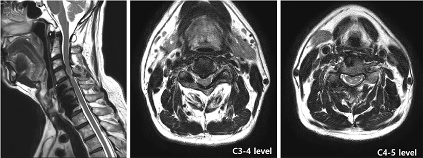
2017년 본 저자들은 CSM에 대한 PCDF 이후 발생한 좌측 편마비 사례를 경험하였다. 2년 전 제5-6-7경추 간 ACDF를 받았던 63세 남자 환자는 Hoffman 징후를 동반한 우측 제1,2 수지의 저린감을 호소하였다. 영상의학적 검사에서 제4-5경추 레벨에서 척수연화증이 있으며, 제3-4 경추 간 추간판탈출증으로 척수가 압박되는 소견이 확인되어 본 저자들은 제3-4-5경추 간 PCDF를 계획하고 수술을 진행하였다(Fig. 1).

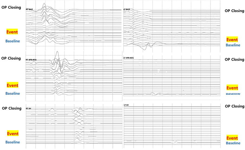
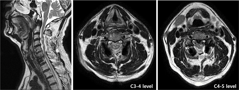
제3-4-5경추의 후궁과 후관절 경계부위를 절삭하고, 좌측에서 부드럽게 들어올리는 과정에서 척수가 부풀어오르는 것을 확인하였다. 바로 확인한 MEPs 감시에서는 진폭이 낮아지거나 소실되었고, 뒤 이어 양측 하지의 SSEPs 또한 진폭이 감소하였다. 집도의는 척수의 재관류 손상을 의심하여 평균동맥압을 95 mmHg 이상으로 유지하고 추가적으로 제2경추의 상부 후궁절제술(upper laminectomy) 및 제6경추의 하부 후궁절제술(lower laminectomy)을 시행하였다. 이후 우측에서 기록한 MEPs는 기저 수준까지 회복이 되었으나, 좌측에서 기록한 MEPs는 완전 소실되어 변화가 없었다. 이어서 제3,4,5경추의 외측괴로 나사못을 고정하고, 제3-4-5경추 간 유합술을 완료하였다. 우측에서 자극한 SSEPs는 회복되었으나, 좌측에서 자극한 SSEPs는 회복되지 않았고, 좌측 상하지의 MEPs는 완전 소실된 상태로 수술이 종료되었다(Fig. 2). 환자는 수술 이후 극심한 좌측 통증 및 0/5의 근력의 좌측 편마비를 호 소하였고, 수술 직후 촬영한 경추 T2강조 자기공명영상에서 제3-4경추 레벨에서 좌측으로 척수부종을 동반한 고신호강도가 확인되었다(Fig. 3).


2018년 Antwi 등은 CSM에 대한 PCDF 이후 재관류손상으로 인해 좌측 편마비가 발생한 사례를 보고하였다[31]. 저자들은 제4-7경추에서 후궁절제술을 통한 후방 감압 및 제3–7경추에서 유합술을 진행하였는데, 수술 중 상근막 조직(suprafascial tissues)의 봉합 이후, 좌측 상하지의 MEPs가 완전 소실되어 추가적으로 제7경추 후궁절제술을 하였다. 그러나 좌측의 MEPs는 회복되지 않은 상태로 수술이 종료되었다.
2020년 Mathkour 등은 PCDF 이후 우측 편마비가 악화된 사례를 수술에 따른 재관류손상으로 보고하였는데, 후궁절제술을 통한 제3-5경추의 후방감압 및 제2-6경추 간 유합술을 하고, 근막을 봉합하고 있는 중 MEPs의 변화는 없었으나, SSEPs의 진폭이 50% 이상 저하되었다[34].
2019년 Papaioannou 등은 PCDF 이후 사지마비가 발생한 사례를 보고하였다[35]. 저자들은 2년 전 제4-6경추 간 ACDF를 받았던 79세 남자 환자에서, 제3-6경추 간 후궁절제술을 통한 후방 감압 및 제2-7경추 간 유합술을 시행하였다. 수술 중 MEPs 및 SSEPs 감시에서 유의한 변화는 관찰되지 않았으나, 수술 직후 촬영한 경추 자기공명영상에서 제6-7경추 레벨에서 T2강조 고신호강도가 확인되었다.
2019년 Wiginton 등은 제1경추 후궁절제술 이후 발생한 사지마비 사례를 보고하였다[6]. 제1경추의 후궁절제술 직후 양측 상지의 SSEPs가 사라지며 점차 양측 하지의 SSEPs 도 완전히 소실되었고, 양측 상지의 MEPs 도 유발되지 않아, 추가적으로 제2경추 후궁 상부의 부분절제술을 시행하였고, 이후 SSEPs와 MEPs는 회복되기 시작했다.
2020년 Busack 등은 CSM 환자의 PCDF 이후 발생한 사지마비 1례를 재관류손상에 따른 후유증으로 보고하였는데, 저자들은 제3-6경추에서 후궁절제술을 마친 이후 수술 시야에서 척수가 빠르게 부풀어 오르며 확장되는 것을 확인하였고, 이후 양측 상하지의 MEPs가 완전 소실되고 SSEPs는 감소하였다[36].
IONM이 함께 시행된 8사례 중 1사례를 제외하고 MEPs 또는 SSEPs의 변화가 동반되었다. 수술 중 MEPs와 SSEPs가 의의 있게 변화를 보인 경우는 각각 총 6례이며, MEPs와 SSEPs가 함께 변화를 나타낸 경우는 5례이다. 또한 MEPs 또는 SSEPs가 진폭이 저하되거나 소실되는 시점은 근막을 봉합하는 시점이 3례이며, 감압 직후가 4례이다.
경추 감압 직후 척수가 부풀어 오르며 확장되는 사례는 재관류 손상으로 보고된 총 14례 중 3례이다. 감압 직후 IONM의 유의한 변화나 척수의 확장되는 양상은 총 5례가 문헌보고에서 확인된다.
경추 감압 수술 이후 척수 재관류손상의 유병율은 2%–5.7%로 보고되고 있어 이에 병태생리학적 기전은 명확하지 못하다[32,37].
실험동물연구 및 인간에 대한 선행연구에 따르면 대체로 오랫동안 심하게 척수가 압박되었던 싱태에서 감압술을 시행할 경우 재관류가 발생하게 되며, 척수 내 급격하게 혈류량이 늘어나며 즉각적으로 척수의 부종이나 확장이 발생한다[9,30, 31,35,36]. 이러한 급격한 척수의 확장은 혈액-척수 장벽(blood-spinal cord barrier)의 붕괴로 기인할 가능성이 있으며, 혈액-척수 장벽의 붕괴로 인해 TNF-α와 IL-1β 같은 염증매개체(inflammatory mediator)의 척수 내로 통과를 증가시키고, 산화 스트레스(oxidative stress)를 유발할 수 있다[30,34,35,37]. 또한, 산화 스트레스 이후 자유기(free radical)나 특정 인지질 연쇄반응으로 척수 재관류 상태에서 세포손상을 받는다[9,30,38]. 특히 전각세포가 있는 척수 내부의 회색질은 백색질에 비해 혈류가 풍부하여 이러한 변화에 손상을 받기 쉽다[9].
2004년 보고된 연구에 의하면, 경추 후궁성형술을 받은 114명의 환자 중 수술 3주 후 T2강조 자기공명영상에서 7명(6.1%)에서 척수내 고신호강도를 보이는 부종소견을 보였고, 이 중 4명은 신경학적 결손 소견을 나타났으며, 신경학적 결손을 호소한 9명 중 5명에서 척수부종을 동반한 T2강조 고신호강도가 확인된다고 한다[39]. 척수의 재관류손상의 유병률이 좀 더 높을 수 있으나, 경추 감압 수술 이후 신경학적 결손이 드물게 발생하기에 CSM에 대한 수술에서 척수의 재관류손상이 발생하는 위험 요인을 고려해 보아야 한다.
이러한 위험 요인으로 산화 염증 반응이 가능한 고령, 만성고혈압 환자에서 나타날 가능성이 있다고 제시하고 있으나, 척수의 재관류 손상으로 보고된 14례 중 65세 이상인 경우는 3례에 불과하여, 다른 위험 요인도 고려해 보아야 한다[34,35].
또한 척수의 관류는 뇌척수액의 압력에 의존하기에 수술 중 감압된 부위에서 뇌척수액이 올라가며 수막낭이 부풀어지는 것이 쉽게 관찰되어 뇌척수액의 압력을 조절하는 기능 또한 위험요인의 하나로 제시되고 있다[36].
실험동물연구에 따르면 후궁절제술 이후 척수가 노출된 부위로 척수혈류(spinal cord blood flow)가 약 30%–200% 증가하는데, 대체로 회색질(gray matter)로 혈류량이 급격하게 증가하여 약 90분간 지속된다[40]. 또, 다른 실험동물연구에서는 척추 감압(spinal compression) 이후 충혈(congestion)이 보고된 경우는 적으나, 증등도 이상의 척수 손상에서는 척추 감압 후 15분 이내로 충혈현상이 보인다고 하며, 또한 인간을 대상으로 한 연구에서는 압박된 척수가 감압되면 유의하게 척수의 관류가 증가되었다고 하며, 척추 수술 이후 척수의 부종이 발생하면 48–72시간 정도 유지될 수 있다고 한다[37,41].
그리고 경추 압박성척수병에 대한 경추 후궁성형술 79례에 대한 IONM의 연구에서 13례에서 IONM의 변화(MEPs의 변화 12례 및 SSEPs의 변화 4례)를 보였다. 저자들은 IONM의 변화를 보이는 경우와 그렇지 않은 경우를 회귀분석을 해보았는데, IONM의 변화는 최대 압박 부위(maximal compressive level)에서 Spinal canal 넓이와 ventral compressive lesion 넓이의 비율에 유의하게 연관되게 나타났으며, IONM의 번화는 모두 부분 절제된 후궁을 들어올리거나, 후궁절제술을 한 직후 나타났다[10].
따라서, 압박된 척수의 정도와 그 부위에서 감압하는 것을 CSM 환자에서 경추 감압시 척수 재관류손상의 위험요인으로 고려해 볼 수 있다.
신경근척수증이 유발된 CSM 환자에서 감압수술은 압박되어 허혈된 척수로 혈류의 공급을 증가시키는 역할도 하며, 따라서 IONM의 호전은 기능적으로 회복할 수 있는지를 예측할 수 있다[15,16,18]. 하지만 오랫동안 심하게 척수가 눌려 취약해진 경우, 오히려 감압수술은 척수의 재관류 손상을 유발할 수 있다. 이러한 재관류 손상은 치명적이기에, 수술 전 압박된 척수의 정도 등 재관류 손상이 발생할 위험요인을 충분히 검토하여 주의 깊게 IONM을 수행해야 한다. 경추 감압 수술 중 척수의 재관류 손상이 발생하면, 빠른 시간에 평균동맥압을 85–95 mmHg 이상으로 시작하여 5–7일간 유지하고, 고용량 스테로이드 프로트콜을 시작해야 하며, 추가적인 감압 수술을 통해 유발원인을 교정 및 더 이상의 손상을 예뱡해야 한다[32].
CSM에 대한 경추 감압 수술에서 IONM은 신경전도경로(neural conduction pathway)에서 급성 손상의 정도와 위치를 결정하고, 이에 대한 유발원인을 교정하며, 저혈압과 갑은 급성 전신변화를 신속하게 관찰하고, 빠르고 적절하게 상기와 같은 과정을 가능하게 한다[42].


