Case Report
수술 중 체성감각유발전위감시에서 인터리브자극방식의 임상적 유용성
안상일1
,
이원우2,
황희원2,
김경민1,2,*
Clinical utility of interleave stimulation method in intraoperative somatosensory evoked potentials
Sang-Il Ahn1,
Wonwoo Lee2,
Heewon Hwang2,
Kyung Min Kim1,2,*
Author Information & Copyright ▼
1Department of Neurology, Yongin Severance Hospital, Yonsei University College of Medicine, Yongin, Korea
2Department of Neurology, Severance Hospital, Yonsei University College of Medicine, Seoul, Korea
*Corresponding author: Kyung Min Kim, Department of Neurology, Severance Hospital, Yonsei University College of Medicine, 50-1 Yonsei-ro, Seodaemun-gu, Seoul 03722, Korea, Tel: +82-2-2228-1600, Fax: +82-2-393-0707, E-mail:
[email protected]
© Copyright 2021 Korean Society of Intraoperative Neurophysiological Monitoring. This is an Open-Access article distributed under the terms of the
Creative Commons Attribution Non-Commercial License (http://creativecommons.org/licenses/by-nc/4.0/) which permits
unrestricted non-commercial use, distribution, and reproduction in any
medium, provided the original work is properly cited.
Received: Jun 04, 2021 ; Revised: Jun 26, 2021 ; Accepted: Jun 27, 2021
Published Online: Jun 30, 2021
ABSTRACT
Intraoperative somatosensory evoked potentials (SSEPs) play a role in reducing postoperative complication. The method of obtaining the SSEP of both upper and lower limbs includes not only a method obtained by serially stimulating each limb, but also an interleave method using auto-sequence. We retrospectively reviewed the clinical and intraoperative monitoring records of 15 patients who underwent both upper and lower limbs intraoperative SSEPs at our hospital from March to September 2020. We compared the amplitude, latency, and acquisition time of the results in each patient when stimulated in serial mode and interleave mode, respectively. Interleave mode stimulation showed no difference in latency, greater amplitude, and shorter acquisition time compared to serial mode stimulation. There were no postoperative complications in 15 patients who underwent intraoperative SSEPs with interleave mode stimulation. Interleave stimulation using auto-sequence can be a clinically useful method for monitoring intraoperative SSEPs of both upper and lower limbs.
Keywords: interleave stimulation; intraoperative somatosensory evoked potential; monitoring; intraoperative; neurostimulation; surgery
서론
수술 중 신경계 감시검사(intraoperative neurophysiological monitoring, INM)는 수술과정에서 신경계의 손상을 방지하기 위해 신경생리학적인 검사를 하는 것으로, 수술 중 신경계의 손상을 조기에 감지하거나 수술 중 손상위험이 높은 주요 신경경로의 정확한 위치를 파악하여 수술 후 신경장애의 위험을 최소화하는 것을 목표로 한다. 중추신경과 말초신경의 손상 위험이 있는 모든 수술에 적용이 가능하며, 진단과 치료의 가치에 대해서 널리 인정되고 있다[1–3].
체성감각유발전위검사(somatosensory evoked potential, SSEP)는 체성감각 자극으로 인해 유발된 전위를 측정하는 검사로 수술 중 신경계 감시검사 중에서 가장 널리 이용되는 검사이며, 양측 상하지에서 자극을 주고, 경추 및 감각신경중추 부위에서 기록한다[4]. 이때 반복적인 자극의 평균화를 통해, 파형을 얻고 수술 중 이 파형의 변화로 신경손상을 예측하게 된다. 신경손상은 매우 짧은 시간에 일어날 수 있어서, 정확하면서도 빠르게 이에 대한 확인을 하는 것은 수술 중 신경계 감시에서 중요하다. 따라서, 정확한 파형을 빠르게 얻을 수 있는 자극 인자들에 대한 연구는 이전부터 진행되어 왔다[5,6].
수술 중 양측 상하지의 체성감각유발전위검사를 진행하여 신경로 손상을 보다 면밀하게 진단할 수 있다. 또한, 양측 상하지의 자극의 순서와 평균화의 방식을 기계마다 시리얼(serial) 방식과 인터리브(interleave) 방식으로 제공하고 있는 경우가 있으나, 두 가지 방식의 임상적 유용성에 대한 비교 연구는 많지 않다. 본 연구는 수술 중 양측 상하지 체성감각유발전위검사에서 인터리브 방식으로 진행하였을 때, 전위, 결과값을 얻는데 걸리는 시간을 시리얼 방식과 비교하고, 수술을 진행한 환자들의 수술 후 합병증 여부를 파악하여, 이에 대한 임상적 유용성을 확인하고자 한다.
본론
2020년 3월부터 9월 사이에 수술 중 체성감각유발전위검사를 받은 환자 15명의 후향적 의무기록과 수술 중 시행한 검사 자료를 이용하여 분석하였다. 환자의 인구학적 정보(나이, 성별), 임상적인 정보(질환, 수술명, 수술 중 신경계 감시 방법, 수술 후 합병증)를 확인하고, 수술 중 체성감각유발전위검사는 Neuromaster MEE-2000 Intraoperative Monitoring System; Nihon Kohden을 이용하여 진행하였고, 시행한 체성감각유발전위검사의 결과는 기존에 사용하였던 시리얼 방식과 인터리브 방식을 사용하였을 때, 각 환자에서 결과도출시간, 전위를 확인하여 비교하였다. 일반적인 수술 중 체성감각유발전위검사 과정에서 수술 환자의 마취 직후에 검사의 정확도 확인을 위해, 수술 중에 시리얼 방식 한차례 확인하고 다음에 인터리브 방식의 체성감각유발전위검사를 진행하는 과정에서 얻어진 결과를 이용하였다.
전체 15명의 환자의 인구학적 특성, 임상정보, 수술 중 체성감각유발전위의 변화, 수술 후 합병증은 Table 1에 나타내었다. 환자들은 평균 59세(연령범위 42–74세), 10명이 남성, 5명이 여성이었다. 경추 수술 9명, 뇌동맥류 수술 5명, 뇌수막종 수술을 1명이 받았으며, 수술 중 체성감각유발전위검사의 변화가 확인된 경우가 3명에서 있었고, 퇴원시에 합병증은 확인되지 않았다.
Table 1.
Demographic and clinical characteristics of patients with intraoperative somatosensory evoked potentials
| No |
Sex |
Age |
Diagnosis |
Operation |
Intraoperative SEP change |
Post-op complications |
| 1 |
M |
47 |
Herniated cervical disc |
Anterior spinal fusion |
None |
None |
| 2 |
M |
74 |
OPLL |
Cervical laminoplasty |
All, amp 50%↓lat 10%↑ |
None |
| 3 |
M |
62 |
Cervical myelopathy |
Anterior spinal fusion |
None |
None |
| 4 |
M |
54 |
Cervical myelopathy |
Posterior spinal fusion |
None |
None |
| 5 |
M |
64 |
Meningioma |
Craniotomy of removal |
None |
None |
| 6 |
M |
66 |
Cervical myelopathy |
Anterior spinal fusion |
None |
None |
| 7 |
M |
73 |
Herniated cervical disc |
Anterior spinal fusion |
None |
None |
| 8 |
F |
55 |
Cerebral aneurysm |
Clipping with aneurysm |
None |
None |
| 9 |
M |
42 |
Cerebral aneurysm |
Clipping with aneurysm |
None |
None |
| 10 |
M |
48 |
Cervical myelopathy |
Anterior spinal fusion |
RL, amp 50%↓lat 10%↑ |
None |
| 11 |
F |
56 |
Cerebral aneurysm |
Clipping with aneurysm |
None |
None |
| 12 |
M |
74 |
Cerebral aneurysm |
Clipping with aneurysm |
LU, amp 50%↓(short) |
None |
| 13 |
F |
66 |
Cervical myelopathy |
Anterior spinal fusion |
None |
None |
| 14 |
F |
58 |
Cerebral aneurysm |
Clipping with aneurysm |
None |
None |
| 15 |
F |
44 |
Cervical myelopathy |
Anterior spinal fusion |
None |
None |
Download Excel Table
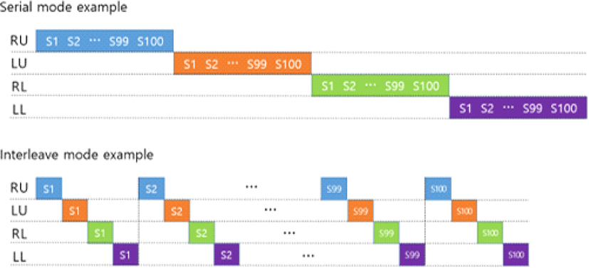
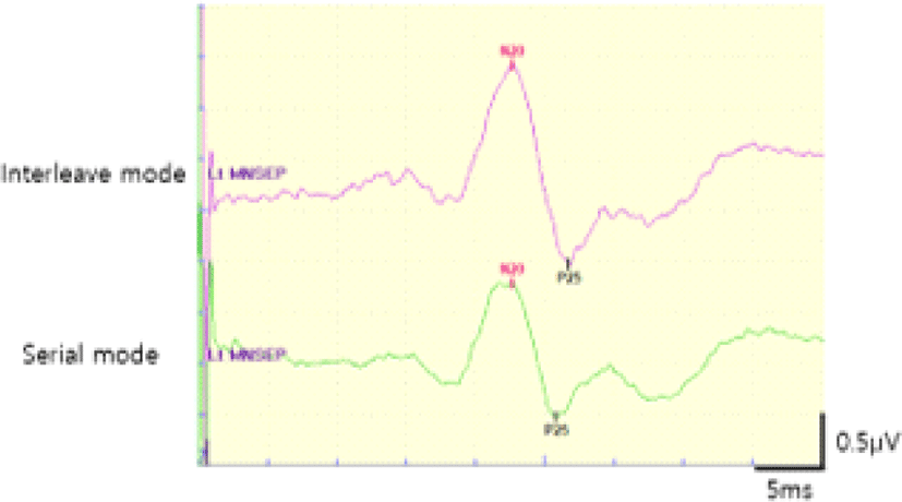
시리얼 자극과 인터리브 자극의 차이는 Fig. 1에 나타내었다. 시리얼 자극이 사지에서 한 부위를 여러 번 자극해서 파형을 얻고, 다른 부위로 넘어가는 것과 다르게 인터리브 자극 방식은 사지를 한 번씩 자극하고, 다음에 다시 사지를 한 번씩 자극하는 식으로 넘어가면서 파형을 얻게 된다. 기존에 사용하던 방식으로 20 mA의 자극을 100번의 평균화를 통해 파형을 얻었으며, 이 때 자극빈도는 시리얼 자극이 5.1 Hz, 인터리브 자극이 2.17 Hz였다. 인터리브 자극은 양측 상하지를 거의 동시에 자극할 수 있어서, 8.68 Hz(2.17 Hz × 4)로 자극이 가능하여 이에 따른 결과도출시간은 시리얼 자극이 78.43초, 인터리브 자극이 46.08초 소요되었다. Table 1에서 5번 환자의 수술 중 체성감각유발전위검사에서 두 자극 방식의 전위와 잠복기가 포함된 파형의 그림은 Fig. 2에 나타내었다. 인터리브 자극 방식의 파형이 전위가 큰 것을 확인할 수 있었다. 15명 환자들 각각의 시리얼 자극과 인터리브 자극으로 얻은 파형에서 각각의 전위는 Table 2에 나타내었고, 시리얼 자극에 비해 인터리브 자극이 평균 1.3배(시리얼 자극 평균 2.45 uV, 인터리브 자극 평균 3.20 uV, p=0.001, Wilcoxon signed-rank test) 전위가 크게 나타나는 것을 확인할 수 있었다.
Fig. 1.
Serial mode and interleave mode.
RU: right upper limb; LU: left upper limb; RL: right lower limb; U: left lower limb; S: stimulation.
Download Original Figure
Fig. 2.
Intraoperative somatosensory evoked potentials with interleave mode and serial mode in patient #5 (left median nerve stimulation).
Download Original Figure
Table 2.
Comparison of amplitude according to stimulation methods
| No |
Amplitude (uV) |
| Serial |
Interleave |
Interleave/Serial |
| 1 |
1.46 (0.10)1) |
1.78 (0.03) |
1.22 |
| 2 |
2.38 (0.16) |
2.39 (0.03) |
1.00 |
| 3 |
5.05 (0.13) |
6.07 (0.08) |
1.20 |
| 4 |
3.39 (0.15) |
4.98 (0.22) |
1.47 |
| 5 |
1.34 (0.03) |
1.97 (0.06) |
1.47 |
| 6 |
1.08 (0.10) |
1.61 (0.06) |
1.49 |
| 7 |
2.15 (0.15) |
3.01 (0.02) |
1.40 |
| 8 |
1.63 (0.10) |
2.24 (0.13) |
1.37 |
| 9 |
1.02 (0.08) |
1.48 (0.06) |
1.45 |
| 10 |
3.02 (0.10) |
4.09 (0.07) |
1.35 |
| 11 |
2.89 (0.11) |
3.84 (0.16) |
1.33 |
| 12 |
3.87 (0.16) |
4.2 (0.03) |
1.09 |
| 13 |
2.85 (0.15) |
3.86 (0.13) |
1.35 |
| 14 |
3.03 (0.07) |
3.91 (0.06) |
1.29 |
| 15 |
1.73 (0.11) |
2.54 (0.03) |
1.47 |
Download Excel Table
고찰
본 연구에서는 인터리브 자극이 시리얼 자극과 비교할 때, 결과도출시간이 빠르고, 전위가 크다는 것을 확인할 수 있었다. 또한 성별, 연령이 다양한 환자군에서 사용이 가능하고, 척추수술과 동맥류 결찰에도 수술 후 합병증 없이 이용 가능함을 확인할 수 있었다. 따라서, 수술 중 체성감각유발전위검사에서 양측 상하지를 자극하는 경우에 유용하게 사용할 수 있는 방식으로 생각된다.
수술 중에는 긴 시간 동안 연속적으로 평균화 검사를 하는 것이 힘들어, 빠른 검사 시간이 필요하나, 빠른 검사 시간을 위해 평균화의 횟수를 줄이는 경우, 신호대잡음비(signal-to-noise ratio, SNR)가 좋지 않아 전위와 잠복기 확인이 어려워지기 때문에 자극의 빈도와 평균화가 중요한 것으로 알려져 있으며, 이전부터 이에 대한 연구가 진행되어 왔다[5,7]. 여기에 더하여, 본 연구에서는 사지를 자극하는 경우, 시리얼 자극과 인터리브 자극이 이용될 수 있으며, 빠른 시간에 더 큰 전위를 얻을 수 있음을 확인할 수 있었다.
다만, 수술의 종류로 볼 때, 양측 상하지의 체성감각유발전위검사가 꼭 필요없는 경우에는 필요한 부위의 검사만 진행하게 되므로, 모든 수술에서 사용할 수 있는 방식은 아니고, 사지를 모두 자극하고 돌아와서 다시 사지를 자극하는 방식이기 때문에, 자극 빈도를 늘리는 데 한계가 있을 수 있다.
미국 임상신경생리학회의 가이드라인에서도 양측을 자극하는 경우에 현재의 검사기계가 인터리브 방식을 제공하고 있다. 이를 통해 결과를 빠르게 큰 전위를 얻을 수 있음을 언급하고 있으며[8], 이전 연구들에서도 인터리브 방식을 이용하여, 수술 중 체성감각유발전위검사를 진행했음을 기술하였다[9,10]. 또한, 사지를 순서대로 감시하는 것이 아니라 사지의 자극 파형을 수술 중에 동시에 감시를 할 수 있기 때문에, 수술 중에 양측 상하지의 체성감각유발전위가 모두 중요한 수술의 경우에는 특히, 유용하게 사용할 수 있는 자극 방식으로 생각된다.
다만, 본 연구는 다음과 같은 한계가 있다. 1) 사례군 연구로 자극 방식의 비교연구를 통해, 합병증의 발생, 수술 중 이상여부 확인을 하지 못했다. 2) 사례의 숫자가 많지 않고, 각각의 환자들의 진단과 수술부위가 달라서, 일반화하기 어렵다. 3) 시리얼 자극과 인터리브 자극 외에 다양한 자극 방식에 대해서도 최근의 기계에서 제공하고 있는데, 이런 자극에 대해서는 확인하지 못했다.
그럼에도 본 연구에서는 실제 환자에서 시리얼 방식과 인터리브 방식이 사용되었을 때 결과도출 시간, 전위의 차이를 확인하여, 수술 중 체성감각유발전위검사에서 실제로 빠르게 신경손상여부를 확인할 수 있다는 것을 보여주었고, 임상적 특성과 합병증 여부를 확인하여 임상적인 유용성을 확인할 수 있었다. 또한 실제 환자에서 자극의 파형을 비교하여 보여줌으로써 수술장에서 검사자가 차이를 느낄 수 있다는 것을 알리는 것에 의미가 있다고 생각한다. 앞으로 다양한 자극방식의 비교 연구가 이루어진다면 정확하고 빠른 수술 중 신경계 감시가 이루어질 수 있을 것으로 기대된다.
Acknowledgments
This study was supported by a research grant of Yongin Severance Hostpital, Yonsei University College of Medicine, Korea.