Case Report

소뇌교각부종양 수술 중 뇌줄기청각유발검사와 자유근전도를 활용한 전정신경 수술중신경계감시

김홍일1https://orcid.org/0000-0002-5325-0295, 김지현2https://orcid.org/0000-0003-3299-7928, 박진석1,*https://orcid.org/0000-0001-6581-5831
Hongil Kim1https://orcid.org/0000-0002-5325-0295, Jihyun Kim2https://orcid.org/0000-0003-3299-7928, Jinseok Park1,*https://orcid.org/0000-0001-6581-5831
Author Information & Copyright
1한양대병원 신경과
2한양대구리병원 신경과
1Department of Neurology, Hanyang University College of Medicine, Seoul, Korea
2Department of Neurology, Hanyang University Guri Hospital, Hanyang University College of Medicine, Guri, Korea
*Corresponding author: Jinseok Park, Department of Neurology, College of Medicine, Hanyang University, 222 Wangsimni-ro, Seongdong-gu, Seoul 04763, Korea, Tel: +82-2-2290-8368, Fax: +82-2-2296-8370, E-mail: jinspark@hanyang.ac.kr

© Copyright 2022 Korean Society of Intraoperative Neurophysiological Monitoring. This is an Open-Access article distributed under the terms of the Creative Commons Attribution Non-Commercial License (http://creativecommons.org/licenses/by-nc/4.0/) which permits unrestricted non-commercial use, distribution, and reproduction in any medium, provided the original work is properly cited.

Received: Mar 30, 2022 ; Revised: Jun 08, 2022 ; Accepted: Jun 16, 2022

Published Online: Jun 30, 2022

ABSTRACT

Cerebellopontine angle tumors including schwannoma or meningioma are located around the vestibular nerve. In the case of surgical removal, intraoperative neurophysiologic monitoring (IONM) modality such as brainstem auditory evoked potential (BAEP) could detect its neurologic function. We demonstrate 2 cases of cerebellopontine angle surgery using BAEP as an IONM modality. And we also suggest how to deal with the case where BAEP is not measured, by using triggered electromyography.

Keywords: evoked potentials; evoked potentials; auditory; brain stem; intraoperative neurophysiological monitoring

서론

수술중신경계감시는 수술 중 수술 부위에 대한 해부학적 위치를 국소화하고 수술 중 발생할 수 있는 손상을 미리 감지하여 수술 후 발생할 신경학적 합병증에 대한 위험성을 낮추기 위해 시행한다[1].

대표적인 수술중신경계감시 검사는 운동유발전위(motor evoked potential, MEP), 체성감각유발전위(somatosentory evoked potential, SEP), 자유 진행 근전도(free running electromyography)가 있다. 이는 척수수술이나 뇌동맥류 결찰 등 운동신경계나 감각신경계를 주로 감시할 때 유용하며, 청신경과 청각 관련 회로가 포함되거나, 손상될 위험이 있는 수술의 경우 뇌줄기청각유발전위(brainstem auditory evoked potentials, BAEPs)를 추가로 확인한다.

뇌줄기청각유발전위 검사는 반복 청각 자극을 통해 얻는 청각유발전위로 다른 유발전위검사와 달리, 청신경계와 뇌간을 포함하기 때문에, 청신경이나 뇌간종양을 포함하는 후두와 수술에 이용할 수 있다[2,3]. 이에 저자들은 청신경 주위의 소뇌교각부종양 수술에서 뇌줄기청각유발전위검사를 시행한 증례와, 뇌줄기청각유발전위검사에서 파형이 형성되지 않은 경우 간접적으로 얼굴신경 유발근전도(triggered electromyography, triggered EMG)를 사용하여 모니터링한 증례를 보고하고자 한다.

증례

본 증례의 수술에서 사용된 기기는 Medtronics사의 NIM- ECLIPSE NS를 사용하였다. 마취는 프로포폴(propofol), 레미펜타닐(remifentanil)을 사용하여 완전정맥마취(total intravenous anesthesia, TIVA)를 시행하였다. 수술중신경계감시는 운동유발전위, 체성감각운동유발전위, 자유 진행 근전도 및 뇌줄기청각유발전위 검사를 시행하였다. 뇌줄기청각유발전위 검사를 위해 스폰지 이어폰을 통한 교대극성(alternating polarity) 자극방식을 사용하였으며, 파형의 측정을 위해 Cz, A1, A2의 전극을 사용하였다. 또한 수술 중 가능한 빠른 시간 내에 판독 가능한 파형을 얻기 위해서 43.9 Hz의 자극속도와 400회의 자극횟수를 이용하였다[4]. 경고기준(alarm criteria)으로는 The American Clinical Neurophysiology Society의 권고대로 wave V의 1 ms 이상의 잠복기 연장 혹은 50% 이상의 진폭 감소를 기준으로 하였다[5].

증례 1

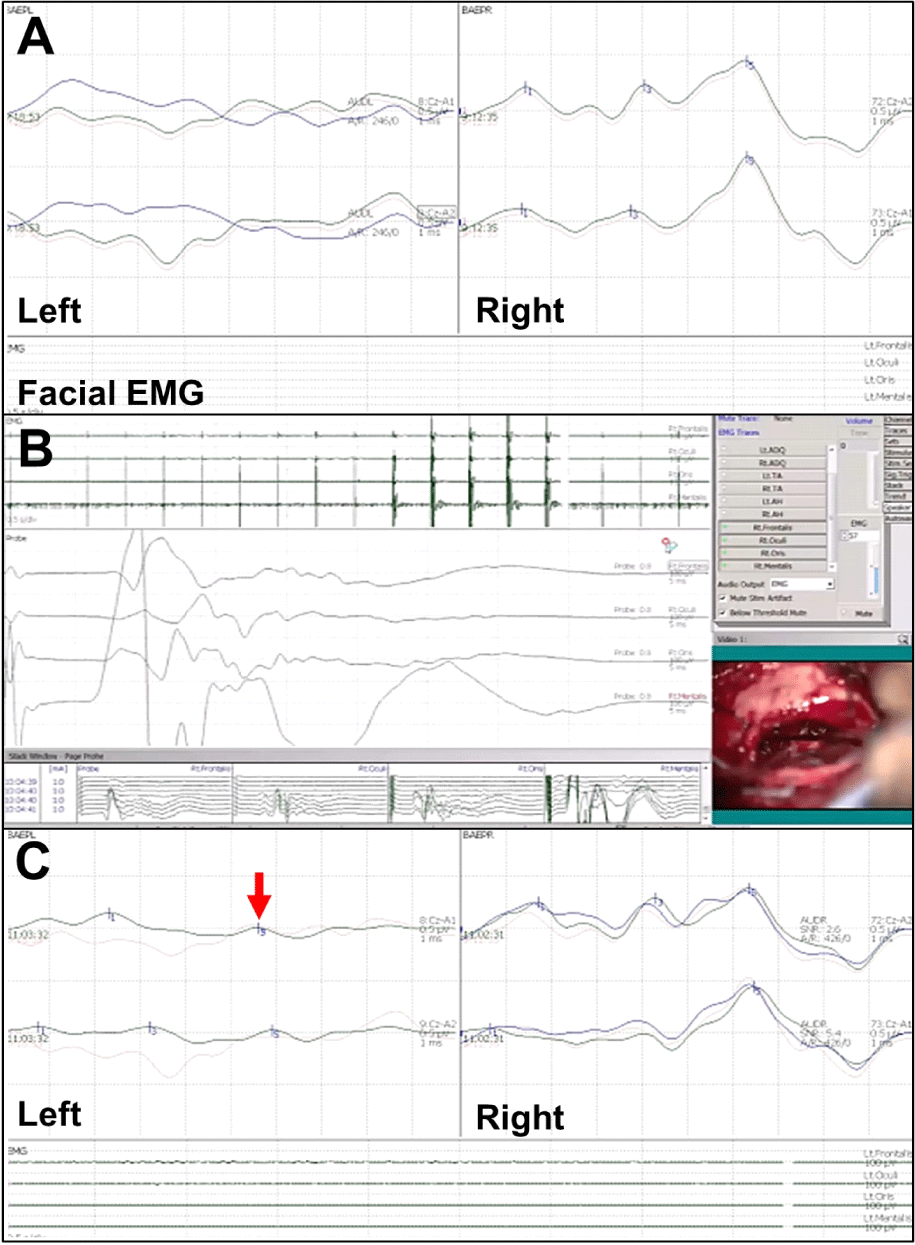
57세 여자 환자가 약 1달전부터 발생한 청각이상으로 내원하였다. 환자는 내원 약 5개월 전부터 간헐적인 우측 안면 부위 통증, 2개월 전부터 두통과 비특이적 어지럼증을 호소하였고, 1달전부터 우측 귀에서 울리는 소리가 들리고, 청력이 저하된다고 호소하였다. 고혈압, 당뇨 그리고 대상포진의 과거력을 가지고 있었다. 뇌자기공명영상검사에서, 우측 소뇌교각의 1.7 × 0.5 × 1.0 cm 크기의 뇌수막종이 확인되었다(Fig. 1-A). 상기 소견에 대하여 구불정맥굴뒤접근법을 통한 종양제거술을 시행하였다. 수술중신경계감시로 뇌줄기청각유발전위 검사를 시행하였고, 수술 초기에는 baseline과 비교하여 파형 및 진폭이 정상 범위였으나, 종양제거 중 우측 Wave V 잠복기가 baseline과 비교하여 1 ms 연장되고, 진폭이 50% 이상 감소된 소견이 관찰되었다. 수술을 일시적으로 중단 후 견인을 풀고, warming 한 이후 진폭은 회복되었으나, 잠복기는 연장된 상태로 수술을 종료하였다(Fig. 2). 수술 후 안면 통증과 청각이상은 회복되었으나, 이명과 어지럼증은 지속되었다.

join-4-1-44-g1
Fig. 1. Case 1,2 pre-operative brain MRI. (A) Brain MRI showed meningioma at right cerebellopontine angle. FLAIR view (red arrow). (B) Brain MRI showed acoustic schwannoma at left internal acoustic canal. T2 axial view (red arrow). MRI: magnetic resonance imaging; FLAIR: fluid attenuated inversion recovery.
Download Original Figure
join-4-1-44-g2
Fig. 2. Case 1. Result of intraoperative brainstem auditory evoked potentials (BAEP) and facial electromyography (EMG). (A) Baseline, (B) In left BAEP, amplitude of wave V was decreased during procedure (red arrow). (C) Latency prolongation of wave Vduring procedure (red arrow), (D) Amplitude was recovered after tumor resection.
Download Original Figure
증례 2

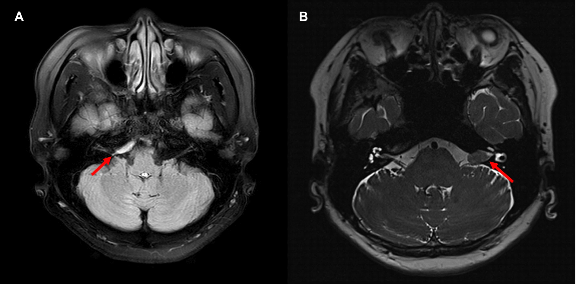
65세 여자 환자로 3년 전부터 좌측 청력저하가 있었으며, 3달 전부터 악화되어 내원하였다. 환자는 고지혈증의 과거력을 가지고 있었다. 뇌자기공명영상에서 좌측 청신경초종이 확인되었다(Fig. 1-B). 수술은 구불정맥굴뒤접근법을 통해 진행되었다. 수술중신경계감시에서 뇌줄기청각유발전위 검사를 시행하였고, baseline부터 좌측의 파형이 형성되지 않았다. 뇌줄기청각유발전위 검사를 통한 수술중 신경계감시가 불가능하여 얼굴신경 유발근전도 시행하였고, 1 mA로 좌측 안면신경을 자극하여 좌측 이마근, 눈둘레근, 입둘레근, 턱끝근의 파형형성여부를 통해 좌측 종양 절제연을 확인하여 제거하였다. 종양 제거 이후 뇌줄기청각유발전위 검사에서 wave I, V가 측정되었다(Fig. 3). 수술 종료 후 환자의 증상은 변화가 없었으나, 2달 후 주관적인 청력저하의 회복이 관찰되었다.

join-4-1-44-g3
Fig. 3. Case 2. Result of intraoperative brainstem auditory evoked potentials (BAEP), Facial Electromyography (EMG), and Triggered EMG. (A) There were poor wave formations of left BAEP on baseline, (B) Triggered EMG was used to excise the tumor instead of using BAEP, (C) BAEP Wave formation was minimally recovered after excision of tumor (red arrow).
Download Original Figure

고찰

본 증례들은 청각 경로에 영향을 주는 종양을 절제하는 수술에 있어 뇌줄기청각유발전위를 이용하여 수술중신경계 감시를 시행한 증례이다. 뇌줄기청각유발전위 검사 값 중 Wave V의 잠복기와 진폭은 수술 후 청력회복에 있어 높은 민감도와 특이도를 갖으므로 이에 대한 모니터링을 중점으로 시행하였다[6].

첫 번째 증례는 수술 중 wave V의 잠복기와 진폭이 유의하게 감소되어 수술자에게 경고하였으며, 이후 진폭이 회복된 상태로 수술을 종료하였고, 두 번째 증례는 뇌줄기청각유발검사에서 wave 형성이 되지 않아 얼굴신경 유발근전도를 사용하여 감시하였고 이후 wave 형성을 확인한 뒤 수술을 종료하였다. 뇌줄기청각유발전위 검사는 청각을 자극할 때 청신경을 거쳐 뇌줄기를 통과하여 머리의 두정에서 발생하는 작은 전위를 기록하는 검사이다. 검사 중 기록되는 각 파형들은 청각신경계의 신경해부학적 경로와 순차적으로 연관되어, 이 파형들을 분석하여 청각신경계의 전도장애 또는 뇌줄기 병변의 문제를 확인할 수 있다. 또한 수술중신경계감시에 이용되는 운동유발전위, 체성감각유발전위 및 근전도 검사와 비교하였을 때, 흡입 마취제와 신경근육차단제의 영향을 상대적으로 덜 받는다[7]. 이러한 특징들로 인해 뇌줄기청각유발전위 검사는 수술 중 청각신경계를 모니터링하는 데 유용하게 사용된다.

뇌줄기청각유발전위 검사는 다른 신경계 감시 중에서도 뇌줄기를 모니터할 수 있다는 장점이 있다. 하지만 증례 2에서 보았듯이, 처음부터 뇌줄기청각유발검사에서 wave 형성이 되지 않는 경우에는 얼굴신경을 모니터링함으로써 간접적으로 청신경계의 손상을 유추해볼 수 있다. 청신경은 얼굴신경 및 중간신경(nervus intermedius)과 함께 하나의 신경 다발(acousticofacial bundle)을 형성하여 소뇌교뇌각부터 내이도까지 나란히 주행한다는 해부학적 특징이 있다[8]. 이에 얼굴신경의 유발근전도(triggered EMG)에서 수술 부위의 주변부에 직접 전기 자극을 가하여 얼굴신경이 지배하는 근육의 반응을 기록하고 이를 통해 수술 중 청각신계의 손상을 유추해 볼 수 있다.

일반적으로 뇌줄기청각유발전위 검사를 시행할 때 초당 8–10회의 속도로 1,000–4,000번의 자극을 준 후 유발되는 전위를 평균화하며 이 경우 상당한 시간이 소요되는데 반해, 본 증례에서 시행한 방법과 같이 자극 횟수 자극 속도를 조절할 경우 짧은 시간에 검사 시행이 가능하였다.

이외에도 전기와우청력검사(electrocochleography)를 활용하여, 청각 자극 후 청신경에서 직접 얻을 수 있는 CNAP (Cochlear nerve action potentials)의 파형을 기록함으로써 수술 후 발생할 수 있는 청각 기능의 손실을 최소화할 수 있다. 최근 2 cm 이상의 크기가 큰 청신경초종에서 육안적으로 청신경을 확인하기 어려운 경우에도 Cotton electrode를 이용하여 청신경의 지속적인 모니터링이 가능하게 되어, 수술 중 뇌줄기청각유발전위 검사 및 얼굴신경의 유발근전도 검사와 함께 활용 시 뇌신경계에 대해 다방면으로 감시를 할 수 있어 유용할 것으로 생각된다[9].

수술중신경계감시를 위해 운동유발전위, 체성감각유발전위, 자유진행 근전도 검사를 이용한다. 그러나 본 증례와 같이 수술 중 특정한 뇌신경계의 기능을 감시하기 위해서는 해당 신경계의 해부학적 특징을 고려하여 보다 유연하고 다양한 접근이 필요하다.

Ethical approval

This article does not require IRB/IACUC approval because there are no human and animal participants.

Conflicts of interest

No potential conflict of interest relevant to this article was reported.

References

1.

Korean Society of Intraoperative Neurophysiological Monitoring, Korean Neurological Association, Korean Academy of Rehabilitation Medicine, Korean Society of Clinical Neurophysiology, Korean Society of Electromyography and Electrodiagnostic Medicine. Clinical practice guidelines for intraoperative neurophysiological monitoring: 2020 update. J Intraoper Neurophysiol. 2020;2(1):1-10.

2.

Park KD. Brain stem auditory evoked potential. Ann Clin Neurophysiol. 2001;3(2):187-90.

3.

Seo DW. Intraoperative neuromonitoring. Ann Clin Neurophysiol. 2008;10(1):1-12.

4.

Joo BE, Park SK, Cho KR, Kong DS, Seo DW, Park K. Real-time intraoperative monitoring of brainstem auditory evoked potentials during microvascular decompression for hemifacial spasm. J Neurosurg. 2016;125(5):1061-7.

5.

Guideline 11 C: recommended standards for intraoperative monitoring of auditory evoked potentials. Milwaukee, WI: American Clinical Neurophysiology Society; 2009.

6.

Lepski G, Arévalo A, Roser F, Liebsch M, Tatagiba M. Electrophysiological predictors of hearing deterioration based on AEP monitoring during petroclival meningioma resection. Neurosurg Rev. 2020;44(3): 1601-9.

7.

Legatt AD. Mechanisms of intraoperative brainstem auditory evoked potential changes. J Clin Neurophysiol. 2002;19(5):396-408.

8.

Burmeister HP, Baltzer PA, Dietzel M, Krumbein I, Bitter T, Schrott-Fischer A, et al. Identification of the nervus intermedius using 3T MR imaging. Am J Neuroradiol. 2011;32(3):460-4.

9.

Ishikawa M, Kojima A, Terao S, Nagai M, Kusaka G, Naritaka H. Cochlear nerve action potential monitoring for preserving function of an unseen cochlear nerve in vestibular schwannoma surgery. World Neurosurg. 2017;106:1057.e1-7.