Case Report

경피적 바늘 삽입법을 이용한 수술 중 외안뇌신경 감시의 유용성: 증례

추승호1,https://orcid.org/0000-0001-6956-6350, 조현진1,https://orcid.org/0000-0001-9563-1849, 김동엽2, 송주연1https://orcid.org/0000-0003-2640-2107, 김재림1https://orcid.org/0000-0002-3404-199X, 김용균1, 배효은1, 김동준1https://orcid.org/0000-0002-2306-0757, 서대원1,*https://orcid.org/0000-0002-9266-9355
Seung Ho Choo1,https://orcid.org/0000-0001-6956-6350, Hyunjin Jo1,https://orcid.org/0000-0001-9563-1849, Dongyeop Kim2, Jooyeon Song1https://orcid.org/0000-0003-2640-2107, Jae Rim Kim1https://orcid.org/0000-0002-3404-199X, Young-Kyun Kim1, Hyoeun Bae1, Dong Jun Kim1https://orcid.org/0000-0002-2306-0757, Dae-Won Seo1,*https://orcid.org/0000-0002-9266-9355
Author Information & Copyright
1성균관대학교 의과대학 삼성서울병원 신경과
2이화여자대학교 의과대학 이대서울병원 신경과
1Department of Neurology, Samsung Medical Center, Sungkyunkwan University School of Medicine, Seoul, Korea
2Department of Neurology, Ewha Womans University Seoul Hospital, Ewha Womans University College of Medicine, Seoul, Korea
*Corresponding author: Dae-Won Seo, Department of Neurology, Samsung Medical Center, Sungkyunkwan University School of Medicine, Seoul 16419, Korea. Tel: +82-2-3410-3599, Fax: +82-2-3410-0052, E-mail: daewon@skku.edu

These authors contributed equally to this work.

© Copyright 2022 Korean Society of Intraoperative Neurophysiological Monitoring. This is an Open-Access article distributed under the terms of the Creative Commons Attribution Non-Commercial License (http://creativecommons.org/licenses/by-nc/4.0/) which permits unrestricted non-commercial use, distribution, and reproduction in any medium, provided the original work is properly cited.

Received: Apr 06, 2022 ; Revised: Jun 17, 2022 ; Accepted: Jun 20, 2022

Published Online: Jun 30, 2022

ABSTRACT

Intraoperative neurophysiologic monitoring (INM) is effective method to prevent operation-related nerve damage in various kinds of neurosurgery. In skull base surgery, there is a possibility of iatrogenic damage to extraocular cranial nerves (EOCNs), which can be prevented with proper INM. We report a case with chondroma involving clivus and compressing pons who had intraoperative EOCN monitoring using the percutaneous needle insertion method during extended endoscopic transsphenoidal approach for the resection of the tumor. During the tumor removal, the structure suspected of being the right abducens nerve was exposed, and it was confirmed that electromyography activity was generated from the lateral rectus muscle by applying electrical stimulation to this structure. Postoperatively, the patient had no diplopia. Our case supports that intraoperative EOCN monitoring using the percutaneous needle insertion could be useful for the assessment of EOCN function during skull base surgery.

Keywords: electromyography; intraoperative neurophysiologic monitoring; EOCNs

서론

안구운동을 담당하는 외안근(extraocular muscles, EOMs)은 3번 뇌신경인 눈돌림신경(oculomotor nerve), 4번 뇌신경인 도르래신경(trochlear nerve), 및 6번 뇌신경인 외전신경(abducens nerve)의 지배를 받는다. 해면정맥동(cavernous sinus, CS), 위안와틈새(superior orbital fissure) 또는 추체경사대부(petroclival region)가 침범되는 두개저 수술(skull base surgery)과 해면정맥동 수술(cavernous sinus surgery), 그리고 뇌전증 치료를 위한 측두엽절제술 중 이러한 외안뇌신경(extraocular cranial nerves, EOCNs)이 손상되기 쉽다[15]. 수술 중 외안뇌신경의 손상은 수술 후 복시를 유발할 수 있고, 이는 종종 단안 시력(monocular vision)을 필요로 하고 입체 시력(stereoscopic vision) 상실로 이어지며, 주변 시야, 이차 약시(secondary amblyopia) 및 기능성 실명의 위험을 초래할 수 있기 때문에 환자의 삶의 질에 심각한 영향을 미칠 수 있다[6,7]. 그러므로 해부학적으로 외안뇌신경과 연관 있는 수술에서 외안뇌신경을 보존하는 것은 수술 과정에서의 주요 고려 사항이다.

두개저 수술을 예로, 수술 중 신경계 감시가 시행되지 않은 두개저 수술에서는 의인성 뇌신경 손상의 발생률이 14%–68%로 보고되었고[8,9], 신경계 감시 방법을 사용했던 경우에는 뇌신경 손상이 2%–47%로 보고되었다[8,1012]. 따라서 외안뇌신경에 대한 수술 중 신경계 감시는 수술 중 뇌신경의 손상을 예측 및 예방할 수 있다[1316].

안구운동을 제어하는 외안근은 외안뇌신경에 의해 신경지배되기 때문에 수술 중 외안뇌신경을 모니터링하는 데 근전도(electromyography, EMG)를 사용할 수 있다. 수술 중 외안뇌신경 감시 방법으로, 외안근 자체에 전극을 직접 적용하는 결막경유기법(transconjunctival technique)이 개발되었다. 이 방법은 외안근 자체에 직접 전극을 적용할 수 있어 감시의 정확도를 높일 수는 있으나, 침습적 방법이고 안과의의 도움 없이는 수행할 수 없으며, 대략 1시간이 소모된다는 단점이 있다[3,7,17]. 이러한 방법을 대체하기 위해, 최근 비침습적이며 손기법(free hand technique)으로 수행 가능한 경피적 바늘 삽입 방법을 통한 감시법이 소개되었다[18].

저자들은 경접형동접근법(transsphenoidal approach, TSA)을 통한 두개저 종양 제거 수술 중, 손기법으로 경피적 바늘 삽입 방법을 통해 수술 중 외안뇌신경을 모니터링한 증례를 보고하고자 한다.

증례

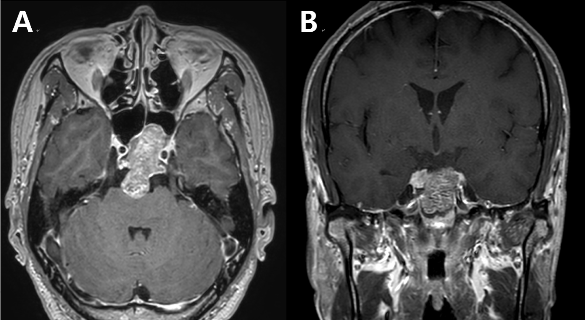
남자 45세 환자가 내원 2주 전부터 발생한 일과성 좌측 상, 하지 마비 증상을 주소로 내원하였다. 환자의 좌측 상, 하지 마비 증세(MRC grade 3)는 약 1–2분간 지속되었다가 완전히 호전되었고, 혀가 굳어져 말이 잘 안 나오는 증상도 동반되었다고 하였다. 이러한 증상이 총 10회 정도 반복되어 타원에서 뇌 MRI(magnetic resonance imaging)를 시행하였고, 경사대부(clivus)를 침범하며 접형동 쪽으로 돌출되고 전교조(prepontine cistern)로도 두개내 침범하여서 뇌교를 압박하고 있는 4.4 cm 크기의 조영 증강되는 종양이 관찰되었다(Fig. 1). 신경학적 검진 상 내원 당시에는 상, 하지 근력의 손상 및 구음 장애뿐만 아니라 외안뇌신경 관련한 신경학적 진찰 상에서 이상 소견은 관찰되지 않았다. 환자는 뇌 MRI 상 관찰된 종양에 대해 확장된 내시경적 경접형동접근법(extended endoscopic transsphenoidal approach)으로 종양 제거술을 시행하기로 하였다.
join-4-1-48-g1
Fig. 1. Brain magnetic resonance imaging of the patient: axial view (A) and coronal view (B). There is a 4.4 cm sized skull base chondroma involving clivus, sphenoid sinus, and intracranial extension to compress pons.
Download Original Figure

마취는 프로포폴(propofol)과 레미펜타닐(remifentanil)을 사용하여 완전정맥마취(total intravenous anesthesia)로 시행하였다. 기관삽관을 위한 근이완제는 로쿠로늄(rocuronium) 50 mg을 1회 정맥주사하였고, 이후 수술 종료 시까지 투여하지 않았다. 수술 중 신경계감시는(Xltek Protektor 32, Natus Medical, Middleton, WI, USA) 장비를 이용해, 운동유발전위(motor evoked potentials, MEP), 체성감각유발전위(somatosensory evoked potentials, SSEP)를 시행하였다. 운동유발전위는 상지에서 짧은 엄지 벌림근(abductor pollicis brevis), 새끼손가락 벌림근(abductor digiti minimi), 하지에서 앞 정강근(tibialis anterior), 엄지발가락 벌림근(abductor hallucis)에서 시행하였고, 체성감각유발전위는 정중신경(median nerve), 후경골신경(posterior tibial nerve)에서 시행하였다. 추가적으로 눈돌림신경, 도르래신경 그리고 외전신경에 의해 지배되는 외안근에 대해 자유 진행 근전도(free-running electromyography, fEMG)와 유발 근전도(triggered electromyography, tEMG)를 시행하였다. 눈돌림신경 감시(oculomotor nerve monitoring)를 위해 하직근(inferior rectus), 내직근(medial rectus), 도르래신경 감시(trochlear nerve monitoring)를 위해 상사근(superior oblique), 외전신경 감시(abducens nerve monitoring을 위해 외직근에서 근전도 반응을 확인하였다. 외안근 기록 전극(EOM recording electrodes)으로는 길이 13 mm, 지름 0.4 mm의 피하바늘전극(subdermal needle electorode) (Xi’an Friendship Medical Electronics, Shanxi, China)을 사용하였다. 바늘전극(needle electrodes)은 외안근에 삽입하기 전에 직각으로 구부려 준비하였고(Fig. 2-A), 안구의 손상을 줄이기 위해, 왼손으로 안구를 외안근으로부터 멀어지게 밀어내면서 바늘전극을 안와 가장자리에 수직으로 삽입하였다(Fig. 2-B). 내직근을 타겟으로 하는 바늘 전극은 6시 방향의 하안와(inferior orbital rim) 가장자리에 수직으로 삽입하였고, 내직근을 타겟으로 하는 바늘 전극은 3시 방향(오른쪽 눈), 상사근을 타겟으로 하는 바늘은 10시 방향(오른쪽 눈), 외직근을 타겟으로 하는 바늘은 9시 방향(오른쪽 눈)에 외측 안와(lateral orbital rim) 가장자리에 수직으로 삽입되었고 바늘 끝은 안와 측벽을 따라(along the lateral wall of the orbit) 삽입하였다(Fig. 2-C). 기준기록전극(reference recording; cathode)과 활성기록(active recording; anode)은 약 5 mm 간격을 두고 위치시켰고, 접지 전극(ground electrode)은 반대쪽 허벅지에 적용하였다. 저주파 필터(low-freqeuncy filter)는 30 Hz, 고주파 필터(high-frequency filter)는 3,000 Hz, notch filter 60 Hz로 설정하였고, 시간축(time base)은 100 ms/division, 민감도(sensitivity)는 70 μV로 세팅하였다. 외안뇌신경에 단극성(monopolar) 항전류성(constant current) 자극을 주고 외안근에서 근전도 반응을 관찰하였으며, 자극의 빈도 및 세기는 4.7 Hz, 1.0 mA로 하였다.

join-4-1-48-g2
Fig. 2. Subdermal needle electrodes are percutaneously inserted into the extraocular muscles with the free-hand technique. (A) Bending electrodes before insertion into the extraocular rectus muscles. (B) Electrode insertion into the lateral rectus muscle with the sharp edge perpendicular to the lateral orbital rim at 9 o’clock through the skin. (C) Overview prior to surgical draping. Yellow and orange electrodes inserted into medial rectus, white and blue electrodes inserted into superior oblique, grey and red electrodes inserted into inferior rectus, black and violet electrodes inserted into lateral rectus.
Download Original Figure

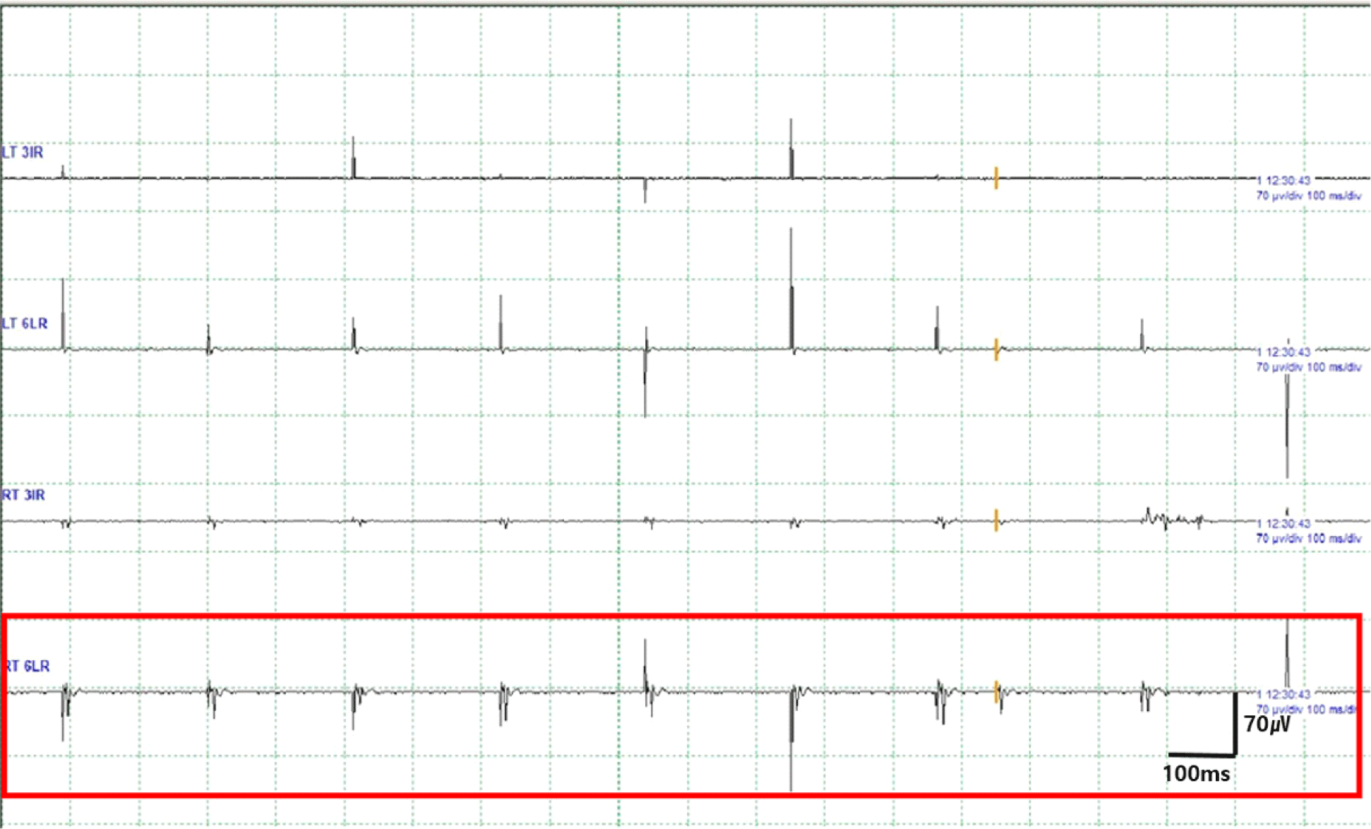
종양을 절반 이상 제거하였을 때 오른쪽 6번 뇌신경으로 의심되는 구조물이 노출되었고, 이 구조물에 직접 전기 자극을 가했을 때 오른쪽 외직근에서 근전도 활동전위가 나오는 것을 확인하였다(Fig. 3). 이후 유발 근전도를 통해 오른쪽 6번 뇌신경의 주행을 확인한 뒤, 이 신경을 보존하면서 추가적으로 종양 제거를 시행하였다. 종양을 모두 제거한 후에도 오른쪽 6번 뇌신경을 자극했을 때 오른쪽 외직근에서 근전도 활성이 잘 유지됨을 확인함으로써, 수술 중 오른쪽 6번 뇌신경의 기능이 잘 보존되었음을 예측할 수 있었다. 수술 중 운동유발전위(MEP), 체성감각유발전위(SSEP) 감시 검사에서도 파형의 유의한 변화는 관찰되지 않았다.
join-4-1-48-g3
Fig. 3. Triggered EMG demonstrates well evoked EMG activities (in red box). EMG: electromyography.
Download Original Figure

종양의 병리 소견은 연골종(chondroma)이었고, 수술 직후 환자는 복시 등의 신경학적 이상을 호소하지 않았으며, 수술 후 시행한 뇌 MRI 추적검사에서 잔존 종양의 확인되지 않았다.

고찰

수술 중 육안으로 정확히 구분되지 않는 종양 주위의 신경의 위치를 확인하기 위하여 근전도 검사를 이용할 수 있다. 근전도 검사는 뇌신경에 의해 지배를 받는 근육에 피하바늘전극(subdermal needle electrode)을 부착을 하여 파형분석을 하며, 수술 중 신경손상이 발생할 때마다 바로 감지가 가능한 자유진행 근전도 방법과 직접 신경을 전기 자극하여 신경과 뇌종양을 구별하는 방법(유발 근전도)이 있다. 특히 유발 근전도는 수술 중 신경의 위치를 확인하는 데 도움을 주고, 그 부위를 피해서 종양을 절제할 수 있어 수술 중 신경 손상의 발생을 줄일 수 있다.

두개저 수술에서 외안뇌신경을 보존하는 것이 중요하고, 수술 중 신경계 감시가 외안뇌신경을 보존하는 데 유용하다는 보고들이 있어왔지만, 아직까지 외안근에 대한 수술 중 근전도 모니터링은 일상적으로 수행되지는 않았다. 이는 주로 기술적인 어려움 때문이었다. 외안근은 안와강(orbital cavity) 깊숙이 있어 접근하기 어렵고, 작고 얇으며(외안근 운동단위(motor unit) 사지 골격근 운동단위에 비해 10–40배 작다), 바늘 전극의 움직임으로 인한 안구(globe)의 우발적 천공 및 안와 내 출혈(intraorbital bleeding)의 위험을 수반한다[6,19].

외안근에 정확히 전극을 위치시키기 위해 외안근 자체에 전극을 직접 적용하는 결막경유기법이 개발되었다. 그러나 이러한 방법은 매우 침습적이고 안과의의 도움 없이는 수행할 수 없으며, 대략 1시간이 소모된다[3,7,17].

그에 비해서 본 증례처럼 바늘 전극을 손기법으로 외안근에 경피적으로 삽입하는 방법은 시술의 위험성이 적고 시간이 적게 걸리는 장점이 있다. 환자의 결막하 출혈(subconjunctival hemorrhage)은 바늘을 삽입할 때 안구 결막의 작은 혈관을 바늘 끝으로 찔러서 발생할 수 있는데, 이 방법을 통한 바늘 삽입 시에는 항상 안구를 반대편으로 밀어내면서 바늘을 삽입하기 때문에 안전성을 높일 수 있다. 또한 이 방법은 검사자가 수행하기 쉽고 15분 내외로 시간이 소모되어 오래 걸리지 않는다. Zi-Yi Li 등은 이 방법을 통한 외안뇌신경의 수술 중 근전도 모니터링이 실현 가능하고 효과적임을 보여주었다[18].

본 증례에서는 기존의 고전적인 결막경유기법에 의한 방법보다 덜 침습적이고 시간이 덜 소요되는 손기법을 통한 경피적 바늘 삽입 방법을 이용한 수술 중 외안뇌신경 감시의 유용성을 확인하고자 하였다. 저자들은 수술 중 유발 근전도 감시를 통해 외전신경의 주행을 확인할 수 있었고, 이를 통해 외전신경을 보존하면서 종양을 제거할 수 있었다. 본 증례에서 사용한 외안뇌신경 감시 방법은 비교적 쉽게 수행 가능하였으며, 덜 침습적 방법이어서 술기 후 합병증 동반 가능성도 적었다.

본 증례에서 사용된 방법에 몇 가지 한계점이 있다. 첫째, 직접 외안근에 바늘을 적용하지 않고 경피적 바늘 삽입 방법을 사용하기 때문에 외안근으로의 정확한 바늘 적용이 안될 수 있다. 둘째, 눈돌림신경 감시를 위해 내직근에 바늘을 적용할 때, 눈물샘의 손상이 동반될 수 있으므로 주의가 필요하다.

상기 언급한 한계점들이 있지만, 본 증례를 통해 수술 중 외안뇌신경의 감시를 통해 수술 후 발생할 수 있는 외안근 마비를 방지할 수 있는 것을 보였다. 비교적 쉽고 덜 침습적인 방식의 외안뇌신경 감시 방법을 통해 외안근 감시가 필요한 다양한 수술들에 폭넓게 적용하여 수술 후 발생할 수 있는 외안근 마비의 발생을 줄일 수 있다.

Ethical approval

Informed consent for publication of the images was obtained from the patient.

Conflicts of interest

No potential conflict of interest relevant to this article was reported.

References

1.

Gao D, Fei Z, Jiang X, Zhang X, Liu W, Fu L, et al. The microsurgical treatment of cranio-orbital tumors assisted by intraoperative electrophysiologic monitoring and neuronavigation. Clin Neurol Neurosurg. 2012;114(7):891-6.

2.

López JR. Neurophysiologic intraoperative monitoring of the oculomotor, trochlear, and abducens nerves. J Clin Neurophysiol. 2011;28(6):543-50.

3.

Sekiya T, Hatayama T, Iwabuchi T, Maeda S. A ring electrode to record extraocular muscle activities during skull base surgery. Acta Neurochir. 1992; 117(1-2):66-9.

4.

Georgiadis I, Kapsalaki EZ, Fountas KN. Temporal lobe resective surgery for medically intractable epilepsy: a review of complications and side effects. Epilepsy Res Treat. 2013;2013:752195.

5.

Newman S. A prospective study of cavernous sinus surgery for meningiomas and resultant common ophthalmic complications (an American Ophthalmological Society thesis). Trans Am Ophthalmol Soc. 2007;105:392-447.

6.

Schlake HP, Goldbrunner R, Siebert M, Behr R, Roosen K. Intra-operative electromyographic monitoring of extra-ocular motor nerves (Nn. III, VI) in skull base surgery. Acta Neurochir (Wien). 2001; 143(3):251-61.

7.

Sekiya T, Hatayama T, Iwabuchi T, Maeda S. Intraoperative recordings of evoked extraocular muscle activities to monitor ocular motor nerve function. Neurosurgery. 1993;32(2):227-35.

8.

Liang SQ, Liang EH, Chen BD, Chen L. Intraoperative oculomotor nerve monitoring during skull base tumor surgery. Zhonghua Yi Xue Za Zhi. 2012; 92(35):2506-8.

9.

Wu B, Liu WD, Chen LY, Huang GF. Application of far lateral craniocervical approach in the microsurgical treatment of the jugular foramen tumors. Zhonghua Wai Ke Za Zhi. 2013;51(1):49-53.

10.

Maurer J, Pelster H, Amedee RG, Mann WJ. Intraoperative monitoring of motor cranial nerves in skull base surgery. Skull Base. 1995;5(3):169-75.

11.

Thirumala PD, Mohanraj SK, Habeych M, Wichman K, Chang YF, Gardner P, et al. Value of free-run electromyographic monitoring of lower cranial nerves in endoscopic endonasal approach to skull base surgeries. J Neurol Surg B Skull Base. 2012;73(4): 236-44.

12.

Sekhar LN, Møller AR. Operative management of tumors involving the cavernous sinus. J Neurosurg. 1986;64(6):879-89.

13.

Thirumala PD, Kassasm AB, Habeych M, Wichman K, Chang YF, Gardner P, et al. Somatosensory evoked potential monitoring during endoscopic endonasal approach to skull base surgery: analysis of observed changes. Neurosurgery. 2011;69(Suppl 1):64- 76.

14.

Romstöck J, Strauss C, Fahlbusch R. Continuous electromyography monitoring of motor cranial nerves during cerebellopontine angle surgery. J Neurosurg. 2000;93(4):586-93.

15.

Schlake HP, Goldbrunner RH, Milewski C, Krauss J, Trautner H, Behr R, et al. Intra-operative electromyographic monitoring of the lower cranial motor nerves (LCN IX–XII) in skull base surgery. Clin Neurol Neurosurg. 2001;103(2):72-82.

16.

Thirumala P, Lai D, Engh J, Habeych M, Crammond D, Balzer J. Predictive value of somatosensory evoked potential monitoring during resection of intraparenchymal and intraventricular tumors using an endoscopic port. J Clin Neurol. 2013;9(4):244-51.

17.

San-juan D, Barges-Coll J, Gómez Amador JL, Díaz MP, Alarcón AV, Escanio E, et al. Intraoperative monitoring of the abducens nerve in extended endonasal endoscopic approach: a pilot study technical report. J Electromyogr Kinesiol. 2014;24(4):558- 64.

18.

Li ZY, Li MC, Liang JT, Bao YH, Chen G, Guo HC, et al. Usefulness of intraoperative electromyographic monitoring of oculomotor and abducens nerves during skull base surgery. Acta Neurochir. 2017; 159(10):1925-37.

19.

Jensen SF. The normal electromyogram from the external ocular muscles. Acta Ophthalmol. 1971; 49(4):615-26.