Review Article

척추수술에서 운동신경계 감시: MEPs(motor evoked potentials) and D-waves

박진석 1 , 2 , * https://orcid.org/0000-0001-6581-5831
Jinseok Park 1 , 2 , * https://orcid.org/0000-0001-6581-5831
Author Information & Copyright
1한양대학교 의과대학 신경과학교실
2한양대학교병원 신경과
1Department of Neurology, College of Medicine, Hanyang University, Seoul, Korea
2Department of Neurology, Hanyang University Hospital, Seoul, Korea
*Corresponding author: Jinseok Park, Department of Neurology, College of Medicine, Hanyang University, Seoul 04763, Korea, Tel: +82-2-2290-8367, Fax: +82-2-2296-8370, E-mail: Jinspark@hanyang.ac.kr

© Copyright 2025 Korean Society of Intraoperative Neurophysiological Monitoring. This is an Open-Access article distributed under the terms of the Creative Commons Attribution Non-Commercial License (http://creativecommons.org/licenses/by-nc/4.0/) which permits unrestricted non-commercial use, distribution, and reproduction in any medium, provided the original work is properly cited.

Received: Nov 11, 2024 ; Revised: Dec 30, 2024 ; Accepted: Jan 08, 2025

Published Online: Jun 30, 2025

ABSTRACT

Intraoperative neurophysiological monitoring plays an important role in preventing nerve injury and improving the safety of spinal surgery. Among various methods, motor evoked potential (MEP) and D-wave monitoring are essential for assessing motor pathway integrity in real time. This review discusses the mechanisms, technical aspects, advantages, and limitations of MEP and D-wave in spinal surgery.

Keywords: D-wave; evoked potentials, motor (MEP); intraoperative neurophysiological monitoring (IONM); motor system; spinal surgery

서론

수술중신경계감시(intraoperative neurophysiologic monitoring, IONM)는 수술 중 신경계 기능을 실시간으로 감시하여, 수술 중 예기치 못하게 발생할 수 있는 손상을 예방 및 최소화하기 위해 시행되고 있다. 신경손상은 비가역적으로 알려져 있으나, 수술 중 여러 신경계 감시 방법을 통해 비가역적 손상이 발생하기 전 조기 발견하여 대처하는 데 그 목적이 있다. 신경계 감시는 감각신경, 운동신경, 그리고 반사신경계를 감시하는 여러 방법이 제안되어 사용되고 있다. IONM은 뇌수술, 척추 수술, 비뇨생식기계, 말초신경을 포함한 정형외과적 수술에서 폭넓게 활용될 수 있다. 특히 상대적으로 수술의 빈도가 많은 척추 수술 시 유용한 신경계감시 방법으로서 활용된다. 척수(spinal cord)는 뇌신경계의 신경로가 좁은 공간에 밀집되어 있기 때문에, 작은 부위의 손상에도 광범위한 신경기능의 손상이 일어날 수 있어 IONM의 역할이 중요하다. 척추수술(spine surgery)은 척추(vertebra) 부위 감압, 안정 및 고정을 위한 수술과 척수(intra/extramedullary)의 종양과 같은 이상을 수술하는 척수수술(spinal cord surgery)로 분류할 수 있다. 척추 수술의 IONM에 사용되는 대표적인 방법으로 감각신경계를 감시하는 체성감각유발전위(somatosensory evoked potential)와 운동신경계를 감시하는 운동유발전위(motor evoked potential, MEP)가 있다. 그리고 근전도(electromyograph, EMG)는 근육의 반응을 감시하기 위한 자발근전도(free-running electromyography, EMG)와 척추수술 중 삽입된 나사못에 자극을 주어 반응을 평가하는 유발근전도(triggered EMG) 검사를 사용한다. 또한 척수내종양수술에 척수지도화(spinal cord mapping) 방법을 활용하기도 하고, 비뇨생식반사를 활용한 구해면체근반사(bulbocavernous reflex)를 활용한 IONM을 시행하기도 한다[1].

본 종설에서는 척추 수술(spine surgery)의 IONM 방법 중 운동신경계 감시에서 대표적인 운동유발전위(MEP)와 D-파형(D-wave)을 중심으로 살펴보고자 하며, 현재까지의 최신 지견을 함께 논의해 보려 한다.

본론

운동신경계는 척수에서 피질척수로(corticospinal tract)를 통해서 신호가 전달된다. 피질척수로는 가쪽피질척수로(lateral corticospinal tract)와 안쪽피질척수로(medial corticospinal tract)로 나뉘며, 가쪽피질척수로가 전체의 약 90%, 안쪽피질척수로가 약 10%로 구성되어 있다. 가쪽피질척수로는 연수부위의 신경섬유교차(pyramidal decussation)를 통해 신체의 반대쪽 근육 운동을 조절하고, 안쪽피질척수로는 신경섬유교차를 하지 않고 신체 중심부(axial) 근육의 운동기능을 조절한다.

IONM에서는 뇌의 운동피질(primary motor cortex)에서 시작된 신호가 연수부위 신경섬유교차를 통해 척수의 피질척수로, 말초신경, 그리고 근육에 이르는 경로를 감시한다. 이 중 가장 보편적으로 사용되는 것이 근육에서의 운동유발전위(myogenic MEP)이며, 척수에서 신호를 기록하는 방법이 D-파형이다(Fig. 1).

join-7-1-1-g1
그림 1. Schematic diagram of motor evoked potentials (MEPs).
Download Original Figure
1. 운동유발전위(motor evoked potential, MEP)

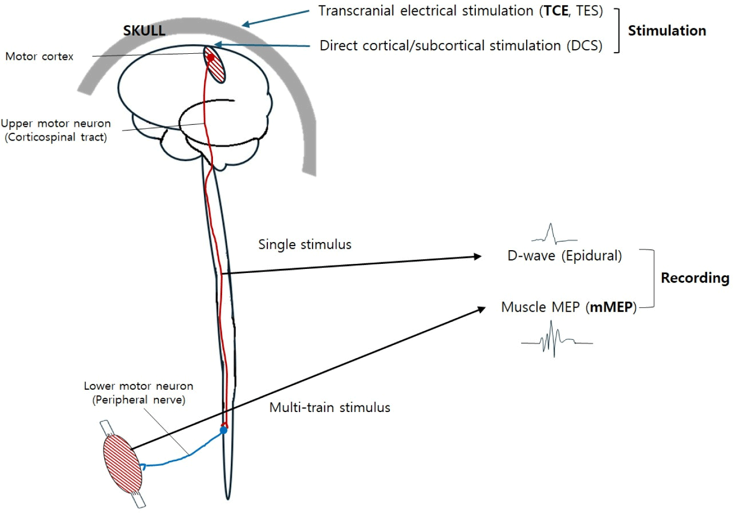
척추 수술 IONM에서 운동신경계를 감시하는 방법 중 가장 보편적인 것이 근육 MEP이다. 운동피질은 경두개전기자극(transcranial electrical stimulation, TES or TCE) 이후 근육에서 기록되는 복합운동활동전위를 기록하는 방법으로, 이는 대뇌피질, 척수, 말초신경, 신경근접합부, 그리고 근육에 이르는 정보를 포함한다. 전기자극은 international 10–20 system의 C3/C4 위치를 400 V 정도의 양극성(anodal) 자극을 사용한다. 경두개전기자극을 하는 여러가지 montage 중, C3/C4가 가장 보편적이며, 보다 넓고 깊은 자극을 줄 수 있다. C1/2 montage는 C3/4보다 자극의 깊이가 얕고 편측만 자극되어 C3/4 위치가 보편적으로 사용된다[2].

MEP의 자극은 역치를 넘어서는 자극을 주어야 하기 때문에 단일자극(single pulse stimulation)으로는 신호가 전달되지 않는다. 5–7회의 다중자극(multipulse stimulation) 방법을 사용해야 자극 역치가 낮아져 신호가 전달된다고 알려져 있다[3]. 다중자극 방법에서 각 자극의 지속시간은 0.5 ms 미만이 추천된다[4]. 다중자극에서 자극과 자극 사이의 간격(interstimulation interval)은 너무 길면 연접 후 전위중첩이 소실되고, 너무 짧으면 불응기(refractory period)에 들어가게 되어 피질척수로는 효과적으로 자극하지 못한다[5]. 자극의 세기(intensity)는 일반적으로 400 V 정도이며, 화상, 경련 등의 부작용을 예방하기 위해 에너지의 총합이 저항이 1 kΩ일 때 50 mJ을 넘지 않아야 한다[2,6]. 일반적으로 TES로 인한 부작용의 비율은 매우 낮으며, 전기자극으로 인한 경련의 발생률은 15,000명을 TES-MEP 감시를 시행했을 때 5명에서 관찰되었다[3]. 4,179명을 대상으로 한 연구에서는 0.8%만이 경련을 보였으며, 척추 수술에서의 발생률은 0.2%였다[7]. 이 밖에 전기자극부위의 탈모, 치아파절, 혀의 열상 등이 보고되어 수술 준비 및 IONM 중 주의가 필요하다[8].

근육 MEP의 기록은 양측 사지의 근육을 선택하여야 하며, 주로 피하에 삽입하는 전극(subdermal needle electrode)을 사용한다. 가장 보편적으로 선택되는 근육은 상지에서는 짧은엄지굽힘근(abductor pollicis brevis), 새끼벌림근(adbuctor digiti minimi) 하지에서는 앞정강근(tibialis anterior), 엄지벌림근(abductor hallucis)이다. 또한 수술부위가 상부경추부위나 하부뇌간 부위일 경우 삼각근(deltoid), 등세모근(trapezius)과 같은 근위부 근육을 추가로 선택하고, 하부요추부나 천추부 수술의 경우 항문조임근(anal sphincter)을 선택하기도 한다. 하지의 근위부 근육은 가쪽넓은근(vastus lateralis)을 선택하는데, 흉추부 수술 시 추가하기도 한다. 기록 전극은 수술 중 탈락되지 않도록 피부부위에 밀착하여 정리하도록 주의하여야 한다.

IONM은 MEP를 사용하지만 검사실이 아닌 수술실 환경에서 시행하므로, 검사의 참조치(reference value)가 없으며, 수술시작 시의 기준 파형(baseline)과의 비교를 통해 이상 유무를 판별한다. 그러므로 본격적인 IONM 전에 기준 파형 획득에 주의를 기울여야 한다. 다만 수술 초반에는 진정 및 기관 삽관 시 사용한 마취제 및 근이완제(neuromuscular blocker, NMB)의 영향을 많이 받기 때문에 이 점을 고려해야 한다. IONM에서는 정맥마취(total intravenous anesthesia, TIVA) 방법을 사용하는데 이는 흡입가스마취가 중추신경계의 신경전달을 저해하기 때문이다[9,10]. 신경전달 시 억제작용을 하는 GABA(gamma-aminobutyric acid)의 기능을 활성화시키고, NMDA와 AMPA 수용체를 통한 신경연접 후 흥분성자극을 억제하는 역할을 한다[10]. 또한 뇌파 진폭 및 주파수의 용량의존적 감소를 유발한다고 알려져 있다[9]. NMB는 주로 기관 삽관 시 사용하고, 간헐적으로 환자의 움직임이 있을 때 사용하는 경우도 있다. 그러나 NNB는 근육으로의 신경전달을 저해하기 때문에 근육 MEP 기준 파형과의 비교를 방해한다. 따라서 NMB는 마취 시작 이후 사용하지 않거나 가능한 한 적은 용량으로 유지하여 기준 파형과의 비교에서 방해가 되지 않도록 해야 한다. 더불어 환자의 근력도 고려해야 하며, 최소 MRC grade 3 이상인 경우에 유의미한 결과를 얻을 수 있다.

척추 수술에서 근육 MEP의 경고기준(alarm criteria)은 명확하게 합의되지는 않았다. 다만 잠복기는 척추 수술의 근육 MEP에서 유용성이 낮아, 파형의 크기(amplitude) 변화를 기준으로 감시한다[11]. 일반적으로 기준 파형보다 50% 이상의 진폭 감소를 기준으로 정하고 있으나 70%, 80% 이상을 기준으로 삼은 연구도 있다[12,13]. 다만 이는 보다 민감하게 근육 MEP의 이상을 측정할 수 있으나, 감별하기 쉽지 않다. 근육 MEP의 반응을 일으키는 최소자극 에너지를 기반으로 하는 자극역치(threshold)를 사용하는 방법은 역치가 100 V 이상 증가하면 유의미한 변화로 간주한다[14]. 또한 근육 MEP의 특성상 자극 시마다 변화가 있는데 근육 MEP의 기간과 변이성이 저하되는 것을 기준으로 하기도 한다[15].

2. D-파형

D-파형은 대뇌피질 자극 시 피질의 5번째 세포층의 피라미드세포(pyramidal cell)의 자극에 의해 형성되는 파형으로, GABA interneuron에 의한 I-파형과 함께 측정된다[16]. 다만 I-파형은 마취에 의해 억제되고 D-파형이 IONM에 사용된다[17,18]. D-파형은 근육 MEP와 달리 경두개 단일자극으로 척수의 경막외(epidural)에 위치한 기록전극을 통해 기록한다. 이 때문에 신경근접합부에 작용하는 NMB의 영향이 제한적이다. 마취는 근육 MEP에서와 같이 TIVA를 사용하는데 isoflurane과 같은 흡입마취제의 경우대뇌 피질의 혈류증가로 션트(shunt)가 발생하여 피질척수로를 깊게 자극하지 못해 D-wave가 약하게 측정되게 된다[19]. D-파형의 자극은 C1/2나 C3-4 위치에 단일자극을 시행하게 되고 근육 MEP보다 약한 자극을 주게 된다[1921]. 이런 점 때문에 자극시 근육의 움직임으로 지속적인 자극을 하기 어려운 MEP에 비해 적은 자극으로 지속적인 감시를 할 수 있다. 또한 근육을 거치지 않고 척수에서 피질척수로를 기록하기 때문에 D-파형의 크기를 정량적으로 간주하여 비교하기 용이하다. D-파형의 기록은 앞서 언급했듯이 척수의 경막외의 병변 아래쪽에 위치시킨다. 제한적으로는 10번째 흉추 아래쪽의 하위 척수는 감시가 어려우며, 전극이 가운데 위치하므로 좌우 구분이 어렵다[21]. 또한 전극의 위치에 따라 D-파형이 영향을 받을 수 있으며, 척추측만증과 같이 3차원적으로 위치가 바뀌는 경우 27% 정도의 위양성(false-positive)이 보고된다[22]. 또한 척수내종양(intramedullary spinal cord tumor)이나 방사선 척수병(radiation myelopathy)의 경우 신경섬유로 전달되는 신호의 속도가 각각 달라 시간차를 두고 기록되는 말초신경병의 시간적 분산(temporal dispersion)과 같은 신경전도의 비동기화(desynchronization in condution)가 발생하여 D-wave 측정에 제한이 발생한다[19,21].

경고기준은 일반적으로 50% 이상의 진폭감소를 이용하지만, 수술상황을 고려해야 하며, 언급한신경전도의 비동기화나 자세에 따른 D-파형의 위양성이 발생할 수 있으므로 D-파형 단독이 아닌, 근육 MEP의 변화를 함께 고려하여야 한다[16]. Sala는 D-파형과 MEP의 결과를 바탕으로 대처법과 예후를 기술하였는데, D-파형과 MEP가 변화가 없거나 소실된 경우에는 논란의 여지가 없겠으나, D-파형이 50% 이상 줄어들고, MEP가 소실되었다면 수술을 멈추어야 하며, 운동기능의 영구손상의 가능성이 있다고 제시하고 있다. 또한 D-파형이 변화가 없거나 50% 미만으로 변화한 경우라도 MEP가 관찰된다면 예후의 변화가 없겠으나, MEP의 한쪽 혹은 양쪽 소실이 관찰된다면 잠시 수술을 중단하고 혈류개선 등의 대처를 취하여야 하고, 이는 일시적인 근육기능 손상을 반영한다고 기술하고 있다[19].

결론

척추 수술에서 운동신경계 IONM은 수술 중 신경계 손상을 최소화하며, 수술의 안전성을 높이고, 최종적으로 수술 이후 환자의 기능과 삶의 질을 보장하는 데 기여하고 있다. 척추 수술의 특성상 좁은 수술시야, 수술과정 중 환자의 자세변경, 추궁절제술(laminectomy) 등 수술부위 접근이나 환자 상태에 의한 척수 압박 등의 상황을 여러 신경생리학적 방법을 활용하여 위험을 최소화하고 있고, 향후 더 나은 과학적 기법의 도입 및 연구가 이루어져야 할 것이다. 또한 한가지 기법이 아닌 여러가지 기법을 통합적으로 적용하여야 하는 IONM의 특성상, 통합적인 연구 및 가이드 마련의 제시를 위한 노력이 필요할 것이다.

Ethical approval

Not applicable.

Conflicts of interest

No potential conflict of interest relevant to this article was reported.

References

1.

Kim JS, Park KS. Intraoperative neurophysiologic monitoring in the spine surgery. J Intraoper Neurophysiol. 2019;1(2):1-14.

2.

MacDonald DB, Skinner S, Shils J, Yingling C. Intraoperative motor evoked potential monitoring: a position statement by the American Society of Neurophysiological Monitoring. Clin Neurophysiol. 2013;124(12):2291-316.

3.

MacDonald DB. Safety of intraoperative transcranial electrical stimulation motor evoked potential monitoring. J Clin Neurophysiol. 2002;19(5):416-29.

4.

Bartley K, Woodforth IJ, Stephen JPH, Burke D. Corticospinal volleys and compound muscle action potentials produced by repetitive transcranial stimulation during spinal surgery. Clin Neurophysiol. 2002;113(1):78-90.

5.

Novak K, de Camargo AB, Neuwirth M, Kothbauer K, Amassian VE, Deletis V. The refractory period of fast conducting corticospinal tract axons in man and its implications for intraoperative monitoring of motor evoked potentials. Clin Neurophysiol. 2004;115(8):1931-41.

6.

Szelényi A, Kothbauer KF, Deletis V. Transcranial electric stimulation for intraoperative motor evoked potential monitoring: stimulation parameters and electrode montages. Clin Neurophysiol. 2007;118(7): 1586-95.

7.

Ulkatan S, Jaramillo AM, Téllez MJ, Kim J, Deletis V, Seidel K. Incidence of intraoperative seizures during motor evoked potential monitoring in a large cohort of patients undergoing different surgical procedures. J Neurosurg. 2017;126(4):1296-302.

8.

Yoshida G, Imagama S, Kawabata S, Yamada K, Kanchiku T, Fujiwara Y, et al. Adverse events related to transcranial electric stimulation for motor-evoked potential monitoring in high-risk spinal surgery. Spine. 2019;44(20):1435-40.

9.

Sloan TB. Anesthetic effects on electrophysiologic recordings. J Clin Neurophysiol. 1998;15(3):217-26.

10.

Sorrenti V, Cecchetto C, Maschietto M, Fortinguerra S, Buriani A, Vassanelli S. Understanding the effects of anesthesia on cortical electrophysiological recordings: a scoping review. Int J Mol Sci. 2021; 22(3):1286.

11.

Deletis V, Sala F. Intraoperative neurophysiological monitoring of the spinal cord during spinal cord and spine surgery: a review focus on the corticospinal tracts. Clin Neurophysiol. 2008;119(2):248-64.

12.

Langeloo DD, Journée HL, de Kleuver M, Grotenhuis JA. Criteria for transcranial electrical motor evoked potential monitoring during spinal deformity surgery: a review and discussion of the literature. Neurophysiol Clin Clin Neurophysiol. 2007;37(6): 431-9.

13.

Traba A, Romero JP, Arranz B, Vilela C. A new criterion for detection of radiculopathy based on motor evoked potentials and intraoperative nerve root monitoring. Clin Neurophysiol. 2018;129(10): 2075-82.

14.

Calancie B. Intraoperative neuromonitoring and alarm criteria for judging MEP responses to transcranial electric stimulation: the threshold-level method. J Clin Neurophysiol. 2017;34(1):12-21.

15.

Quiñones-Hinojosa A, Lyon R, Zada G, Lamborn KR, Gupta N, Parsa AT, et al. Changes in transcranial motor evoked potentials during intramedullary spinal cord tumor resection correlate with postoperative motor function. Neurosurgery. 2005;56(5):982-93.

16.

Legatt AD, Emerson RG, Epstein CM, MacDonald DB, Deletis V, Bravo RJ, et al. ACNS guideline: transcranial electrical stimulation motor evoked potential monitoring. J Clin Neurophysiol. 2016; 33(1):42-50.

17.

Capone F, Paolucci M, Assenza F, Brunelli N, Ricci L, Florio L, et al. Canonical cortical circuits: current evidence and theoretical implications. Neurosci Neuroecon. 2016;5:1-8.

18.

Di Lazzaro V, Ziemann U. The contribution of transcranial magnetic stimulation in the functional evaluation of microcircuits in human motor cortex. Front Neural Circuits. 2013;7:18.

19.

Nuwer MR. Intraoperative Monitoring of Neural Function. Amsterdam, The Netherland: Elsevier; 2008.

20.

Deletis V. Intraoperative neurophysiology and methodologies used to monitor the functional integrity of the motor system. In: Deletis V, Shils JL, editors. Neurophysiology in neurosurgery. Amsterdam, The Netherlands: Elsevier; 2002. p. 25-51.

21.

Deletis V. Intraoperative neurophysiology and methodologies used to monitor the functional integrity of the motor system. In: Deletis V, Shils JL, Sala F, Seidel K, editors. Neurophysiology in neurosurgery. 2nd ed. Cambridge, MA: Academic Press; 2020. p. 17-34.

22.

Ulkatan S, Neuwirth M, Bitan F, Minardi C, Kokoszka A, Deletis V. Monitoring of scoliosis surgery with epidurally recorded motor evoked potentials (D wave) revealed false results. Clin Neurophysiol. 2006;117(9):2093-101.