Review Article

수술 중 신경계 감시 방법의 최신 지견

송민근 1 , 2 , * https://orcid.org/0000-0001-8186-5345
Min-Keun Song 1 , 2 , * https://orcid.org/0000-0001-8186-5345
Author Information & Copyright
1전남대학교 의과대학 재활의학교실
2전남대학교병원 재활의학과
1Department of Physical & Rehabilitation Medicine, Chonnam National University Medical School, Gwangju, Korea
2Department of Physical & Rehabilitation Medicine, Chonnam National University Hospital, Gwangju, Korea
*Corresponding author: Min-Keun Song, Department of Physical & Rehabilitation Medicine, Chonnam National University Medical School, Gwangju 61469, Korea, Tel: +82-62-220-5198, Fax: +82-62-228-5975, E-mail: [email protected]

© Copyright 2026 Korean Society of Intraoperative Neurophysiological Monitoring. This is an Open-Access article distributed under the terms of the Creative Commons Attribution Non-Commercial License (http://creativecommons.org/licenses/by-nc/4.0/) which permits unrestricted non-commercial use, distribution, and reproduction in any medium, provided the original work is properly cited.

Received: Mar 12, 2026 ; Revised: Apr 12, 2026 ; Accepted: Apr 16, 2026

Published Online: Jun 30, 2026

ABSTRACT

Intraoperative neurophysiological monitoring (IONM) is an essential component of modern neurosurgical and spinal surgery, enabling real-time assessment of neural pathway function and early detection of neurological injury. Established modalities—including somatosensory evoked potentials, motor evoked potentials (MEP), D-wave, electromyography, and brainstem auditory evoked potentials—have been extensively validated in clinical practice and contribute to reducing the incidence of postoperative neurological deficits. Recent advances have introduced novel monitoring technologies such as quantitative frequency-domain near-infrared spectroscopy, laser speckle contrast imaging, and corticobulbar MEP that complement conventional electrophysiological methods. The integration of artificial intelligence and machine learning algorithms is driving innovation in automated signal interpretation, artifact rejection, and outcome prediction. Emerging domains including wireless high-density electroencephalography, cortical spreading depolarization monitoring, and intraoperative autonomic nervous system monitoring are expanding the scope of IONM. This review comprehensively summarized recent advances in the technical principles and clinical applications of both established and emerging IONM modalities, current advances by surgical subspecialty, and future directions.

Keywords: artificial intelligence; intraoperative neurophysiological monitoring

서론

중추 및 말초 신경계의 기능적 보전은 신경외과 및 척추 수술 후 신경학적 예후를 결정하는 핵심 요인이다. 수술 중 신경계 감시는 신경 경로의 기능을 수술 중 실시간으로 평가하여 비가역적 손상이 발생하기 전에 위험을 조기에 감지하고 교정 조치를 취할 수 있도록 한다[1].

수술 중 신경계 감시의 임상 활용은 1960년대 체성감각유발전위검사를 이용한 척추 수술 감시로 시작되었으며, 이후 운동유발전위검사, 직접 피질 자극 등으로 발전하였다. 이러한 전통적 방법들은 척추측만증 교정, 두개내 종양 절제, 뇌혈관 수술 등에서 신경학적 합병증 발생률 감소에 임상적으로 기여하고 있다[1,2].

최근에는 광학 영상, 다중 감시 통합, 인공지능 기반 분석의 등장으로 수술 중 신경계 감시의 범위가 크게 확장되었다. 수술 중 신경계 감시의 안전성과 유효성은 포괄적 리뷰에서도 확인되었으며[3], PIONEER 컨소시엄과 같은 국제 학술단체들은 수술 중 지도화·감시에 관한 포괄적 가이드라인을 제정하는 등[4] 수술 중 신경계 감시 발전은 국제적으로 가속화되고 있다.

본 종설에서는 전통적 및 최신 수술 중 신경계 감시 방법의 기술적 원리와 임상 적용, 비신경계 수술로의 확장에 대하여 최근 발표 위주로 정리하였으며, 미래 발전 방향을 논의하고자 한다.

전통적 수술 중 신경계 감시 방법

전통적 수술 중 신경계 감시 방법들은 수십 년에 걸친 임상 검증을 통해 신경외과 및 척추 수술 안전성의 근간을 이루고 있다(Table 1). 각 방법은 특정 신경 경로를 표적으로 하여 기능 이상의 조기 감지와 수술 중 의사결정을 지원한다[1].

Table 1. Summary of traditional intraoperative neurophysiological monitoring methods, pathways, and warning criteria
Method Monitoring pathway Indications Warning Criteria
SSEP Posterior column → somatosensory cortex Spinal deformity, aortic surgery, spinal cord tumors ≥ 50% decreased amplitude or ≥ 10% delayed latencies
MEP Motor cortex → corticospinal tract → muscle Spinal cord tumors, scoliosis, cerebrovascular surgery 50%–100% decreased or loss amplitude
D-wave Direct corticospinal tract recording Intramedullary spinal cord tumor resection ≥5 0% decreased amplitude
BAEP Cochlea → auditory cortex (brainstem pathway) Vestibular schwannoma, microvascular decompression Wave V latency ≥ 1 ms delayed or loss
EMG Motor nerve → muscle (neuromuscular function) Facial nerve, spinal nerve root, skull base surgery Sustained firing, reduction of motor unit action potential
NCS/CMAP Peripheral nerve conduction (axon/myelin) Peripheral nerve surgery, spinal cord stimulator implantation Decreased conduction velocity, reduced CMAP
DCS/tcMEP Direct motor cortex stimulation Functional cortex mapping, tumor resection Changes in response threshold, loss of EMG signal
EEG/ECoG Cortical electrical activity (including epileptiform activity) Epilepsy surgery, functional mapping, cerebral ischemia Loss of electrical activity, appearance of epileptiform discharges
VEP Retina → occipital lobe (visual pathway) Visual pathway tumor surgery Delayed latency, decreased amplitude
TCD Transcranial Doppler cerebral blood flow velocity Carotid endarterectomy, cerebrovascular surgery ≥ 50% decreased in blood flow velocity

SSEP: somatosensory evoked potentials; MEP: motor evoked potentials; BAEP: brainstem auditory evoked potentials; EMG: electromyography; NCS: nerve conduction study; CMAP: compound muscle action potential; DCS: direct cortical stimulation; tcMEP: transcranial motor evoked potentials; EEG: electroencephalography; ECoG: electrocorticography; VEP: visual evoked potentials; TCD: transcranial Doppler.

Download Excel Table
1. 체성감각유발전위검사

체성감각유발전위검사는 척수 후주와 대뇌 피질까지의 상행 감각 경로의 기능적 온전성을 평가한다. 정중신경 또는 후경골신경에 전기 자극을 가하고 두피 전극을 통해 피질 반응을 기록하며, 척추 기형 교정 수술, 척수 내 종양 절제, 흉복부 대동맥 수술에서 특히 유용하다. 2024년 미국 신경생리 감시학회(ASNM)는 체성감각유발전위검사 감시에 관한 개정된 입장 성명을 발표하여 체성감각유발전위검사를 감각운동 피질 및 배측주(dorsal column) 위치 파악뿐 아니라 척수·뇌 수술 시 체성감각 경로의 기능 평가를 위한 확립된 감시 양식으로 공식 인정하였으며, 척수 자극기 삽입 최적화, 후두와 종양 제거, 대동맥 수술 등으로 적응증, 경고 기준, 방법론을 갱신하였다. 또한 자극 반복률(stimulation repetition rate)이 증가할수록 피질 반응 진폭이 유의미하게 감소하며, 낮은 반복률 유지가 고품질 피질 반응 확보에 필수적임을 강조하였다. 연속적·다신경 교차자극(continuous interleaved multi-nerve stimulation) 방식이 대부분의 수술 상황에서 가장 우수한 감시 패러다임으로 권고하였다[5]. 소아 특발성 척추측만증 수술에서 다양한 체성감각유발전위검사 변화 유형의 진단적 유용성에 대한 대규모 연구도 보고되었다[6].

2. 운동유발전위검사 및 D파

운동유발전위는 경두개 전기 자극 또는 직접 피질 자극을 이용하여 운동 피질에서 말초 근육까지의 피질척수로를 평가한다. 높은 시간적 분해능으로 운동 경로 손상을 신속하게 감지할 수 있어 척추측만증 수술, 척수 내 종양 절제, 뇌혈관 수술에서 필수적으로 활용된다[1].

경추 감압 수술에서 경두개 운동유발전위의 수술 후 신경학적 결손 예측 능력을 체계적 고찰과 메타분석으로 확인한 연구에서, 경두개 운동유발전위는 손상 예측에 대한 민감도 56%, 특이도 94%, 위험비 19.26으로 검증되었다[7]. 운동유발전위 경고 기준과 가역성에 관한 체계적 고찰도 이루어졌으며[8], 척수 내 종양 절제 시 운동유발전위 단독 대비 체성감각유발전위검사 병용 시 높은 민감도(83.9%, 95% 신뢰구간: 76.8–89.2)를 나타냄과 동시에 높은 특이도(84.2%, 95% 신뢰구간: 66.3–92.3)를 보여 진단적 이점도 확인되었다[9].

D파는 경막외 전극으로 피질척수로의 전기 활동을 직접 기록하는 방법으로, 척수 내 종양 절제 시 근전도 기반 운동유발전위와 상호 보완적으로 활용된다[10]. D파와 근전도 운동유발전위의 진단 정확도를 비교한 메타분석에서, D파는 즉각적, 일시적 및 지속적 운동 결손을 모니터링하는 경우 94.0%, 95.0% 그리고 93.4%의 높은 특이도를 보여 위양성 경보를 줄이는 데 유리하였다[11].

3. 뇌간청각유발전위검사

뇌간청각유발전위검사는 와우에서 청각 피질까지의 청각 경로를 평가하며, 청신경초종 절제 및 미세혈관 감압술에서 특히 중요하다. 파형 V의 잠복기가 1 ms 이상 연장되거나 소실되는 경우 경고 기준으로 삼는다. 2026년 미국신경외과학회에서 청신경초종 수술에서의 뇌신경 감시에 관한 안면·청신경 수술 중 신경계 감시의 역할과 필요성을 다룬 근거기반 가이드라인을 업데이트하였다[12].

4. 근전도 및 반사 기반 감시

근전도는 자발 및 유발 근육 활동을 기록하여 운동 단위와 지배 신경의 기능 상태를 평가한다. 최근에는 전통적 유발전위와 근전도를 넘어, 인간 반사(human reflex) 기반 감시가 수술 중 신경계 감시의 새로운 방향으로 주목받고 있다. Choi 등[13]은 순목반사(blink reflex), H-반사, R1/R2 반사 등 다양한 반사 기반 지표가 특정 신경 구조물의 기능 평가에 효과적임을 보고하였다.

Zhang 등[14]은 미세혈관감압술(microvascular decompression)을 시행한 원발성 반측안면경련 수술에서 순목반사와 측방확산반응(lateral spread response) 병합할수록 예후가 불량할 것으로 예측하는 예측력이 최대 42.86%로 증가함을 보고하였다.

5. 뇌파 기반 감시

표준 뇌전도검사는 전반적 피질 활동을 평가하며, 뇌피질뇌파와 입체 뇌파는 뇌전증 유발 부위와 기능 피질 지도화를 위한 침습적 방법이다. 최근 미국 임상신경생리학회는 수술 중 뇌피질뇌파에 관한 기술 표준을 제정하여 전극 배치, 신호 처리, 해석 기준을 표준화하였다[15].

6. 기타 전통적 방법

수술 중 신경전도검사(nerve conduction study)는 말초 신경 전도 속도와 진폭을 측정하여 축삭 기능을 평가하며, 말초 신경 수술 및 척수 자극기 전극 거치에 활용된다. 직접 피질 자극(direct cortical stimulation)은 운동 피질 지도화와 종양 절제 시 기능 보존에 사용된다. 시각유발전위검사(visual evoked potential)는 시각 경로 종양 수술에서 재조명되고 있으며[16], 경두개 도플러(transcranial Doppler)는 경동맥 내막 절제술 등 뇌혈관 수술에서 최적 혈역학 관리에 기여한다[17].

수술 중 신경계 감시의 최신 기술

의공학, 신호 처리, 계산 신경과학의 발전으로 수술 중 신경계 감시의 범위가 크게 확장되고 있다. 최근 10년간 광학 영상, 인공지능 기반 분석, 무선 기술, 자율신경 감시 등 다양한 혁신이 이루어졌다(Table 2).

Table 2. Recent modalities in intraoperative neurophysiological monitoring technologies and key applications
Technology Principle Clincal applications Advantages
Q-NIRS Frequency-domain near-infrared light absorption analysis Cerebral ischemia detection, aneurysm/AVM surgery Quantitative, non-invasive, continuous measurement
LSCI Blood flow measurement via laser speckle pattern Cortical blood flow mapping, bypass surgery High spatiotemporal resolution, contact-free
Corticobulbar MEP Cranial nerve-innervated muscle EMG after transcranial stimulation Skull base/brainstem tumors, hemifacial spasm surgery Real-time functional assessment of corticobulbar tract
AI/CNN/ML Automated signal pattern recognition via deep learning Simultaneous multi-signal interpretation, automatic ischemia detection Automation, prediction, multimodal integration
SNN-based HFO detection Bio-inspired spiking neural network ECoG-guided resection margin determination in epilepsy surgery Real-time, high specificity, low-power processing
Wireless high-density EEG High-resolution EEG recording via wireless electrode array Broad cortical function monitoring during surgery Real-time cortical mapping without cables
Cortical Spreading Depolarization monitoring Electrode recording of cortical spreading depolarization Cerebrovascular surgery, traumatic brain injury surgery Indicator of progressive ischemic brain injury
Autonomic nervous system monitoring Pelvic/vagus/cavernous nerve EMG/NCS Rectal cancer, prostate, thyroid surgery Functional preservation, improved long-term surgical outcomes

Q-NIRS: quantitative frequency-domain near-infrared spectroscopy; AVM: arteriovenous malformation; LSCI: laser speckle contrast imaging; MEP: motor evoked potentials; EMG: electromyography; AI: artificial intelligence; CNN: convolutional neural network; ML: machine learning; SNN: spiking neural network; HFO: high-frequency oscillations; ECoG: electrocorticography; EEG: electroencephalography; NCS: nerve conduction study.

Download Excel Table
1. 정량 주파수 영역 근적외선 분광법

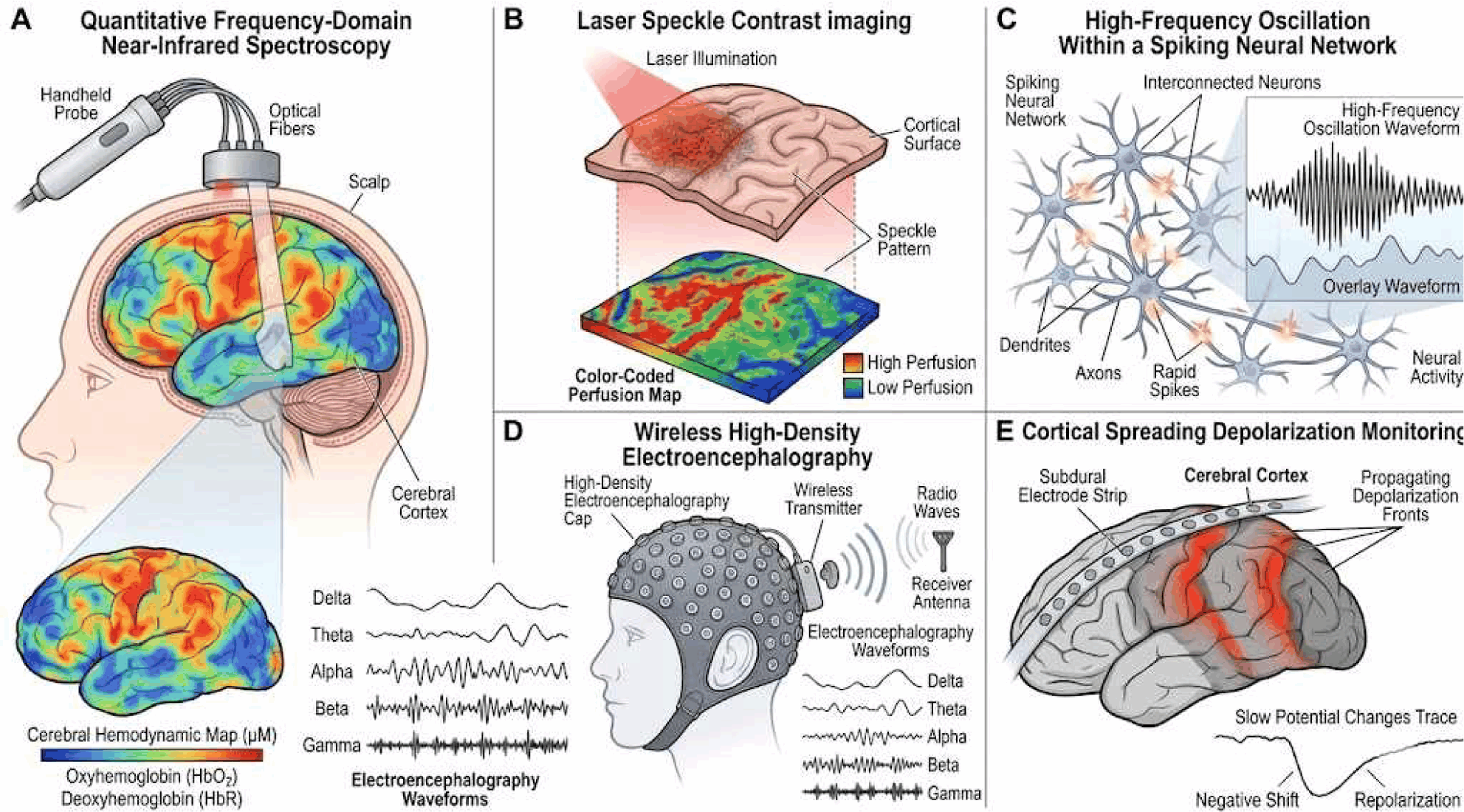
정량 주파수 영역 근적외선 분광법(quantitative near-infrared spectroscopy)은 뇌 조직을 통한 근적외선 광흡수를 분석하여 뇌 산소화와 혈역학을 비침습적·실시간으로 평가한다. 주파수 영역 기법은 조직 산소 포화도와 헤모글로빈 농도를 직접 정량화하며, 동맥류 클립 수술, 뇌동정맥기형 절제, 경동맥 내막 절제술에서 유용성이 입증되었다[18]. 유발전위 파형 변화보다 먼저 관류 결핍을 감지하여 보완적 역할을 한다(Fig. 1-A).
join-8-1-1-g1
Fig. 1. Representative illustrations of emerging intraoperative neurophysiological monitoring (IONM) modalities. (A) Quantitative frequency-domain near-infrared spectroscopy (Q-NIRS), (B) laser speckle contrast imaging (LSCI), (C) high-frequency oscillation within a spiking neural network (HFO within SNN), (D) wireless high‑density electroencephalography, (E) cortical spreading depolarization monitoring.
Download Original Figure
2. 레이저 스페클 대비 영상

레이저 스페클 대비 영상(laser speckle contrast imaging)은 일관성 레이저 광의 스페클 패턴 분석으로 혈류를 지도화하며, 외인성 조영제 없이 높은 시공간 분해능을 제공한다. 피질 관류 감시, 우회 수술 후 혈관 개통성 평가, 혈관 연축 감지에 활용되었다[19]. 비접촉식 특성으로 수술 시야를 방해하지 않는 장점이 있다(Fig. 1-B).

3. 피질뇌간로 운동유발전위

두개저 및 뇌신경 수술에서 피질뇌간로 기능의 실시간 평가는 수술 후 안면 마비, 연하 장애 예방에 중요하다. 경두개 전기 자극 후 뇌신경 지배 근육의 유발 근전도를 기록하는 피질뇌간로 운동유발전위는 두개저 종양, 뇌간 종양, 반안면경련 미세혈관 감압술에 적용된다[20].

소뇌교각부 종양 수술에서 안면 운동유발전위의 예측 가치를 분석한 연구에서, 안면 운동유발전위 소실은 수술 후 안면 기능 결손의 독립적 예측 인자임이 확인되었다(민감도 80%, 특이도 88%)[21]. 안면신경 감시에서는 측면 전파 반응, 안면 F파, 순목반사 등이 상호 보완적으로 활용되며[22] 두개저 수술에서의 안면신경 수술 중 신경계 감시에 관한 체계적 고찰에서도 다중 지표 감시의 중요성이 강조되었다[23].

4. 인공지능 및 기계학습 통합

인공지능 및 기계학습 알고리즘은 복잡한 신경생리학적 데이터의 실시간 해석을 지원한다. 합성곱 신경망, 순환 신경망, 서포트 벡터 머신 등이 유발전위 및 뇌파 신호에 적용되어 허혈성 변화, 발작 활동 감지에서 높은 정확도를 보이고 있으며, 경동맥 내막 절제술에서 뇌전도 기반 합성곱 신경망의 허혈 감지 정확도는 93% 이상으로 보고되었다[24].

인공지능 시스템은 미세한 파형 변화를 자동 감지하여 주관적 해석 의존도를 낮추고 반응 시간을 단축한다. 운동유발전위검사, 체성감각유발전위검사, 정량 주파수 영역 근적외선 분광법, 레이저 스페클 대비 영상 등 다중 데이터 스트림을 통합하여 신경에 대한 포괄적 예측 평가를 가능하게 하며, 임상에서의 인공지능과 인간 판단의 균형에 관한 논의도 활발히 이루어지고 있다[25].

5. 다채널 근전도 데이터셋 및 딥러닝 기반 뇌신경 감시

뇌신경 감시를 위한 인공지능 기반 패턴 인식 개발에서 다채널 근전도 데이터셋 구축이 중요하다. Ma 등[26]은 뇌신경 VII, V, XI, X, XII에서 수술 중 근전도를 동시 기록한 다채널 데이터셋을 제시하였고, 이를 통해 딥러닝 알고리즘이 생리적 활동, 자극 반응, 아티팩트를 구분할 수 있음을 보였다. 이러한 데이터셋은 두개저 및 뇌간 수술의 의사결정 향상에 기여한다.

6. 스파이킹 신경망 기반 고주파 진동 감지

수술 중 뇌피질뇌파에서 고주파 진동(250–500 Hz)은 뇌전증 유발 피질 부위의 생체지표이다. 생물학적 신경 처리에서 영감을 받은 스파이킹 신경망(spiking neural network)은 실시간으로 고주파 진동 사건(high-frequency oscillations)을 신속·저전력으로 분류하여 뇌전증 수술 중 절제 범위의 동적 결정을 가능하게 하였다[27]. 뇌피질뇌파 유도 뇌전증 수술의 체계적 고찰에서는 고주파 진동 기반 절제 시 발작 호전율에 대한 예후 예측은 74%(95% CI 71–77)로 비사용 시에 비해 효과적(OR 2.10, 95% CI 1.37–3.24, p < 0.01)임이 확인되었다[28] (Fig. 1-C).

7. 무선 고밀도 뇌파

최근 무선 고밀도 뇌파(wireless high-density electroencephalography) 시스템이 수술 전후 감시에 도입되었다. 기존 유선 전극 체계와 달리 케이블 없이 실시간 피질 활동을 광역적으로 기록할 수 있어 수술 시야와 마취 관리의 제약을 줄인다[29]. 고밀도 전극은 공간 분해능이 높아 기능적 피질 지도화와 뇌전증 유발 부위 국소화에 유리하다(Fig. 1-D).

8. 피질 확산 탈분극 감시

피질 확산 탈분극(cortical spreading depolarization)은 뇌 허혈·외상 후 2차 뇌손상의 중요한 생리적 지표이다. 경막외 또는 피질 전극을 통해 피질 확산 탈분극을 수술 중 실시간으로 감시함으로써 뇌허혈 진행 여부를 판단하고 신속한 처치를 가능하게 한다[30]. 뇌혈관 수술 및 두부 외상 감압술에서의 임상적 적용이 확대되고 있다(Fig. 1-E).

9. 자율신경 수술 중 신경계 감시

수술 중 신경계 감시의 적용 영역이 신경외과·척추 수술을 넘어 비신경계 수술의 자율신경 보존으로 확대되고 있다. 직장 수술 시 골반 자율신경 감시의 진단 정확도에 관한 체계적 고찰에서 근전도 기반 수술 중 신경계 감시를 통해 수술 후 항문직장 기능장애는 민감도 1.00(95% CI 0.03–1.00), 특이도 0.98(95% CI 0.91–0.99)로 예측되었으며, 배뇨 기능장애 역시 민감도 1.00(95% CI 0.03–1.00), 특이도 0.99(95% CI 0.92–0.99)로 높은 예측 정확도를 보였다[31]. 위 원위부암 수술에서 미주신경 보존과 인도시아닌 그린 라벨링을 병용한 무작위 대조 임상시험(JAMA Surgery [32])에서는 미주신경 보존군에서 0.8%, 담석 발생은 0%로 합병증 발생률이 낮게 관찰되었다. 전립선 근치 절제술에서 해면체 신경의 실시간 지도화 및 감시 기술도 발전하고 있다[33].

수술 분야별 임상 적용

수술 중 신경계 감시의 적용은 수술 분야별 특성, 해부학적 고려 사항, 환자별 위험 인자에 따라 달라지며, 최근 기술 발전으로 각 분야에서의 감시 정밀도와 임상적 유효성이 더욱 향상되고 있다.

1. 두개내 수술
1) 뇌혈관 수술

경동맥 내막 절제술, 동맥류 클리핑, 뇌동정맥기형 절제술에서 체성감각유발전위검사, 운동유발전위, 뇌파의 복합 감시가 표준으로 자리 잡았다. 파열되지 않은 두개내 동맥류의 미세수술 클리핑에서 다양한 수술 중 신경계 감시 방법을 비교한 체계적 고찰과 메타분석에서는 체성감각유발전위검사와 운동유발전위검사 병용 감시가 단일 방법 대비 운동유발전위는 18분 이내, 체성감각유발전위는 16분 이내에 회복될 경우 결손 발생률이 15%였으나, 회복되지 않은 경우 95%까지 증가하여 신경학적 합병증 예측에 우월함이 확인되었다[34]. 정량 주파수 영역 근적외선 분광법과 레이저 스페클 대비 영상은 뇌관류를 실시간으로 평가하여 허혈 조기 감지에 기여한다[18,19].

2) 뇌혈관기형 수술

뇌해면상혈관기형(cavernous malformation) 수술에서 수술 중 신경계 감시의 역할에 관한 서술적 고찰에서는, 체성감각유발전위검사 및 운동유발전위검사가 병변 위치에 따라 선택적으로 적용되어야 하며 혈관기형의 해부학적 특성이 감시 방법 선택에 영향을 미침이 강조되었다[35]. 수면 상태에서의 뇌동정맥기형 절제술에서 수술 중 신경계 감시의 역할에 대한 분석에서도 체성감각유발전위검사/운동유발전위검사의 보완적 활용이 중요하였다[36].

3) 뇌종양 수술 및 기능 보존

기능 부위 인접 신경교종 수술에서 수술 전 지도화와 수술 중 신경계 감시의 통합 전략은 전절제율 향상과 기능 보존을 동시에 달성하는 것으로 나타났다[37]. PIONEER 컨소시엄은 Gerritsen 등[4]에서 신경교종 수술에 관한 포괄적 체계를 제안하며, 수술 중 지도화·감시·의사결정에 대한 국제 권고안을 제시하였다.

언어 기능 지도화 시 직접 전기 자극과 뇌피질뇌파의 비교 연구에서는 직접 전기 자극이 보다 정밀한 언어 부위 동정에 유리한 반면, 뇌피질뇌파는 연속적 광역 감시에 강점이 있어 상호 보완적 활용이 권장된다[38]. 각성 개두술에서의 인지 기능 검사 업데이트에서도 언어·집행 기능 보존을 위한 다양한 평가 프로토콜이 논의되었다[39].

2. 두개저 및 뇌신경 수술

두개저 및 미세혈관 감압술에서 다중 뇌신경 감시가 필수적이다. 안면신경 감시에서는 측면 전파 반응, 안면 F파, 안면 운동유발전위검사, 순목반사 등 다중 지표를 병용하며[22,23], 삼차신경통 미세혈관 감압술에서의 수술 중 신경계 감시 역할에 관한 체계적 고찰에서도 청신경 모니터링을 포함한 다중 감시를 시행한 경우 영구 청력 손실이 0%로 나타난 반면, 비사용군에서는 영구 청력 소실이 발생하여 합병증 예방 효과가 있음을 보고하였다[40].

3. 척추 수술
1) 임상 효과 및 비용 효율성

2025년 발표된 체계적 고찰에서 수술 중 신경계 감시는 척추 수술의 임상 결과 운동유발전위는 가장 높은 민감도(90.2%)를 나타냈고, 체성감각유발전위는 높은 특이도(97.1%)를 보였으며, 경제성 분석에서 신경학적 합병증 발생률이 0.3%를 초과할 시 수술 중 신경계 감시를 시행하는 것이 비용-효과적인 것으로 나타났으며, 증례당 23,000달러 이상의 비용을 절감하는 것으로 보고하였다[41]. 2015–2025년 척추 수술에서 수술 중 신경계 감시 근거를 집대성한 연구에서도 신경학적 합병증 예방 효과가 일관되게 확인되었다[38]. 척추 수술 전반에서 수술 중 신경계 감시의 진단 정확도를 분석한 메타분석에서는 민감도 71%–85%, 특이도 96%–99%로 보고되었다[42].

2) 경추 수술

경추 퇴행성 수술에서 수술 중 신경계 감시가 신경학적 예후에 미치는 영향을 분석한 메타분석에서 수술 중 신경계 감시 적용군에서 수술 후 신경학적 결손 발생률(OR = 0.39 [0.20–0.79])에 대하여 잠재적 보호 효과가 확인되었다[43]. 전방 경추 수술 후 연하 장애 예방을 목적으로 한 전향적 연구에서 수술 중 신경계 감시 적용군에서 비감시군 발생률 43.5% 대비 13.0%로 연하 곤란 발생률이 낮음을 보고하였다[44].

3) 최소 침습 척추 수술

최소 침습 척추 수술(minimally invasive surgery)의 차세대 신경 감시 기술에 관한 보고에서, 소형화된 전극, 무선 근전도, 인공지능 보조 신호 분석이 최소침습 척추 수술 환경에서의 수술 중 신경계 감시 적용을 가능하게 만들었다[45]. 특히 경막 뒤 비교 접근에서 수술 중 신경계 감시의 역할이 확대되고 있다.

4) 척수 내 종양 수술

척수 내 종양 절제 시 D파와 근전도 운동유발전위검사의 상호 보완적 활용이 권장된다[10,11]. 척수 외 경막 내 종양 수술에서 수술 중 신경계 감시 유무에 따른 조기 수술 후 신경학적 결과를 비교한 체계적 고찰에서는 수술 중 신경계 감시 적용군에서 비사용군에 비해 조기 수술 후 신경학적 결손을 유의하게 감소시켰으며(OR 0.39, 95% CI 0.20–0.75; p = 0.005; I2 = 22%), 이는 절대위험감소 9.2%로 보고되어 신경보존에 효과적임을 보고하였다[46].

4. 척수 자극 및 신경 조절 수술

척수 자극기 삽입 시 수술 중 신경계 감시는 전극 거치 정밀도 향상과 신경학적 합병증 예방에 기여한다[47]. 특히 복합근육활동전위(compound muscle action potential)는 체성감각유발전위검사보다 경막외 전극의 중앙선 거치에 더 신뢰할 수 있는 결과를 제공한다. 배근 신경절(dorsal root ganglion) 자극 전극 거치 시 수술 중 신경계 감시에 관한 문헌 고찰 및 증례 연구에서, 배근 신경절 수술에서의 수술 중 신경계 감시 프로토콜이 제안되었다[48].

파킨슨병의 심부 뇌자극(deep brain stimulation) 수술에서 미세전극 기록(microelectrode recording)은 시상하핵의 정밀한 표적 위치 확인에 활용됨을 보고하였다[49].

5. 뇌전증 수술

수술 중 뇌피질뇌파는 뇌전증 유발 부위 확인의 핵심 도구이다. 뇌피질뇌파 유도 뇌전증 수술의 체계적 고찰에서는 뇌피질뇌파 기반 절제 전략이 앞에서 언급한 바와 같이 발작 호전율에 대한 예후 예측은 74%로 고주파 진동 제거가 중요한 예측 인자임이 확인되었다[28]. 미국 임상신경생리학회의 수술 중 뇌피질뇌파 기술 표준(2026) 제정으로 방법론의 표준화가 이루어졌다[15]. 소아 뇌량 절제술에서도 수술 중 신경계 감시의 활용이 보고되었다[18].

6. 비신경계 수술로의 확장

갑상선 수술에서의 되돌이 후두신경 감시는 현재 표준으로 자리 잡았으며[50], 소아 갑상선 수술에서의 수술 중 신경계 감시 패러다임도 정립되고 있다[51]. 식도암 최소 침습 식도 절제술에서 되돌이 후두신경 감시는 성대 마비 발생률 OR 0.24(95% CI 0.13–0.46)로 유의하게 감소시킬 수 있음을 보고하였다[52]. 직장암 수술에서 골반 자율신경 감시[31]와 미주신경 보존 위 수술[32], 전립선 근치 절제술에서 해면체 신경 감시[33] 등으로 수술 중 신경계 감시의 적용 범위가 급속히 확대되고 있다.

수술 중 신경계 감시의 미래 전망

1. 인공지능 강화 감시 및 예측 모델링

인공지능 통합은 자동화된 신호 해석, 아티팩트 제거, 신경학적 예후 예측으로 확장되고 있다. 딥러닝, 특히 스파이킹 신경망을 활용하면 실시간으로 다중 감시 데이터를 분석하여 명시적 파형 변화 이전의 미세 패턴을 포착할 수 있다[24,27]. 임상 수술에서 인공지능과 인간 판단의 최적 균형을 찾는 연구도 진행 중이다[25].

2. 다중 모드 통합 및 개인화 전략

수술 중 신경계 감시의 미래는 전기생리학적 신호를 광학 영상(정량 주파수 영역 근적외선 분광법, 레이저 스페클 대비 영상), 수술 중 초음파, 신경항법 데이터와 결합한 다중 모드 통합에 있다. 유발전위 감시에서의 개인화 접근법을 논한 연구에서는 환자별 기저 신경학적 상태, 해부학적 변이, 수술 위험도에 따른 맞춤형 감시 전략의 중요성이 강조되었다[41].

3. 무선화 및 소형화

무선 고밀도 뇌파 검사와 소형화된 전극 시스템은 수술 환경에서의 제약을 줄이고 실시간 피질 매핑의 범위를 확장한다[29]. WISE 피질 전극과 같은 차세대 기기는 수술 중 신경 감시의 정밀도를 높이는 도구로 주목받고 있다[26].

4. 오류신호 관리 및 신호 품질 향상

마취, 전기 수술 장비, 기계적 간섭에서 비롯되는 오류신호 식별 및 제거는 수술 중 신경계 감시 신뢰성의 핵심 과제이다. 적응형 필터링, 독립 성분 분석, 인공지능 기반 분류를 적용하여 신호 충실도를 향상시키는 연구가 활발히 진행 중이다[18].

5. 비신경계 수술로의 지속적 확장

갑상선, 식도, 직장암, 전립선 등 비신경계 수술로 수술 중 신경계 감시의 적용 영역이 빠르게 확장되고 있다. 자율신경 보존을 위한 감시 기술의 표준화와 임상 근거 축적이 향후 과제로 남아 있으며, 로봇 및 복강경 수술 환경에서의 수술 중 신경계 감시 호환성 개선도 중요한 방향이다.

결론

수술 중 신경계 감시는 제한적 유발전위 측정에서 포괄적 다중 감시 체계로 발전하였다. 체성감각유발전위검사, 운동유발전위검사, D파, 근전도, 뇌간청각유발전위검사 등 전통적 방법은 임상에서 필수적이며, 정량 주파수 영역 근적외선 분광법, 레이저 스페클 대비 영상, 피질뇌간로 운동유발전위검사, 인공지능 기반 신호 분석, 스파이킹 신경망 기반 고주파 진동 감지, 무선 고밀도 뇌파, 자율신경 감시 등 최신 기술들이 수술 중 신경계 감시의 범위와 민감도를 지속적으로 확장하고 있다. 수술 중 신경계 감시의 적용 범위는 비신경계 수술로도 급속히 확장되어, 자율신경 보존이 중요한 갑상선·소화기·비뇨기 수술에서도 신경 기능 보존에 기여하고 있다.

미래의 수술 중 신경계 감시는 손상을 감지할 뿐만 아니라 예측하는 단계로 진화할 것이며, 다중 모드 데이터 스트림의 실시간 통합과 인공지능 기반 의사결정 지원 시스템이 정밀 수술 중 신경계 감시 실현에 핵심 역할을 할 것이다. 이를 위해 임상의, 공학자, 데이터 과학자 간의 지속적인 다학제 간 협력이 요구된다.

Ethical approval

Not applicable.

Conflicts of interest

No potential conflict of interest relevant to this article was reported.

References

1.

Guzzi G, Ricciuti RA, Della Torre A, Turco EL, Lavano A, Longhini F, et al. Intraoperative neurophysiological monitoring in neurosurgery. J Clin Med. 2024;13(10):2966.

2.

Mortazavi SH, Permanoon E, Safarian A, Farboud A. Advancing intraoperative neuromonitoring for complex spinal and nerve preservation: a comprehensive review. J Neurol Sci Res. 2023;3(2):1-6.

3.

Rezapour A, Derakhshani N, Pouyan SN, Alipour V, Ehsanzadeh SJ, Zare-Sadeghi A, et al. Safety and efficacy of intraoperative neuromonitoring: an umbrella review. Health Sci Rep. 2025;8(10):e71370.

4.

Gerritsen JKW, Karschnia P, Bello L, Hervey-Jumper S, Young JS, Seidel K, et al. A comprehensive framework for glioma surgery by the PIONEER Consortium and RANO resect group, part 1: intraoperative recommendations for mapping, monitoring, and decision making. Lancet Oncol. 2026;27(1): E11-23.

5.

Toleikis JR, Pace C, Jahangiri FR, Hemmer LB, Toleikis SC. Intraoperative somatosensory evoked potential (SEP) monitoring: an updated position statement by the American Society of Neurophysiological Monitoring. J Clin Monit Comput. 2024;38(5):1003-42.

6.

Gorijala VK, Reddy RP, Anetakis KM, Balzer J, Crammond DJ, Shandal V, et al. Diagnostic utility of different types of somatosensory evoked potential changes in pediatric idiopathic scoliosis correction surgery. Eur Spine J. 2024;33(4):1644-56.

7.

Reddy RP, Singh-Varma A, Chang R, Vedire A, Anetakis KM, Balzer JR, et al. Transcranial motor evoked potentials as a predictive modality for postoperative deficit in cervical spine decompression surgery: a systematic review and meta-analysis. Glob Spine J. 2024;14(5):1609-28.

8.

Addanki RND, Thomas S, Balasundaram S, Lee BB, Anetakis KM, Balzer JR, et al. Systematic review of alarm criteria and reversibility in motor-evoked potentials during intramedullary spinal cord tumor resection. Clin Neurophysiol. 2025;178:2110966.

9.

Addanki RND, Golish S, Thomas S, McGravey ML, Anetakis KM, Lee BB, et al. Evaluating the diagnostic benefit of adding somatosensory evoked potentials to transcranial motor-evoked potentials compared to either modality alone in intramedullary spinal cord tumor resection: a systematic review and meta-analysis. World Neurosurg. 2025;202: 124360.

10.

Zurita Perea SN, Alvarez Abut PA, Seidel K. A concise guide to D-wave monitoring during intramedullary spinal cord tumour surgery. Medicina. 2024; 60(8):1242.

11.

Thomas S, Addanki RND, Murugan CK, Anetakis K, Lee BB, Balzer JR, et al. A systematic review and meta-analysis to compare the diagnostic accuracy of direct-waves and myogenic motor-evoked potential neuromonitoring during intramedullary spinal cord tumor resection. Oper Neurosurg. 2025;30(4): 513-24.

12.

Patel NS, Carlson ML, Sughrue ME, Olson JJ. Congress of Neurological Surgeons systematic review and evidence-based guidelines update for the role of intraoperative cranial nerve monitoring in the management of patients with vestibular schwannomas. Neurosurgery. 2026;98(2):288-92.

13.

Choi J, Díaz-Baamonde A, Sánchez Roldán MÁ, Pescador AM, Kim JS, Téllez MJ, et al. Advancing intraoperative neurophysiological monitoring with human reflexes. J Clin Neurol. 2024;20(2):119-30.

14.

Zhang Y, Xia L, Lin J, Zhang Y. Retrospective study of the combined application of the blink reflex and lateral spread reflex in microvascular decompression of facial nerve surgery. Ann Palliat Med. 2021;10(12):12310-8.

15.

Schuele S, Husain A, Tatum W, Gavvala J, Selioutski O, Sinha SR, et al. American Clinical Neurophysiology Society technical standards for performing intraoperative electrocorticography. J Clin Neurophysiol. 2026;43(2):101-9.

16.

Olmsted ZT, Silverstein JW, Einstein EH, Sowulewski J, Nelson P, Boockvar JA, et al. Evolution of flash visual evoked potentials to monitor visual pathway integrity during tumor resection: illustrative cases and literature review. Neurosurg Rev. 2023;46(1):46.

17.

Hakimi R, Alexandrov AV, Garami Z. Neuro-ultrasonography. Neurol Clin. 2020;38(1):215-29.

18.

Calderon-Arnulphi M, Alaraj A, Amin-Hanjani S, Mantulin WW, Polzonetti CM, Gratton E, et al. Detection of cerebral ischemia in neurovascular surgery using quantitative frequency-domain near- infrared spectroscopy. J Neurosurg. 2007;106(2): 283-90.

19.

Konovalov A, Grebenev F, Stavtsev D, Kozlov I, Gadjiagaev V, Piavchenko G, et al. Real-time laser speckle contrast imaging for intraoperative neurovascular blood flow assessment. Sci Rep. 2024;14(1): 1735.

20.

Dong CCJ, MacDonald DB, Akagami R, Westerberg B, Alkhani A, Kanaan I, et al. Intraoperative facial motor evoked potential monitoring with transcranial electrical stimulation during skull base surgery. Clin Neurophysiol. 2005;116(3):588-96.

21.

Pan SY, Holdefer RN, Wu HL, Li CR, Guo L. The predictive value of intraoperative facial motor evoked potentials in cerebellopontine angle tumor surgery. Clin Neurophysiol. 2024;166:176-90.

22.

Joo BE. Recent update of intraoperative neurophysiological monitoring during microvascular decompression surgery for hemifacial spasm. J Intraoper Neurophysiol. 2024;6(1):1-7.

23.

Zachem TJ, Bello A, Lee B, Luo E, Woo J, Yoo S, et al. Current state of intraoperative neuromonitoring of the facial nerve during skull base surgery: a systematic review. J Clin Neurosci. 2025;140: 111525.

24.

Hindi R, Pappas G. Unleashing the power of AI for intraoperative neuromonitoring during carotid endarterectomy. Electronics. 2024;13(22):4542.

25.

Parduzi Q, Wermelinger J, Koller SD, Sariyar M, Schneider U, Raabe A, et al. Explainable AI for intraoperative motor-evoked potential muscle classification in neurosurgery: bicentric retrospective study. J Med Internet Res. 2025;27:e63937.

26.

Ma W, Chen L, Pang X, Zou Y. A multichannel electromyography dataset for continuous intraoperative neurophysiological monitoring of cranial nerve. Data Brief. 2024;53:110250.

27.

Burelo K, Sharifshazileh M, Krayenbühl N, Ramantani G, Indiveri G, Sarnthein J. A spiking neural network (SNN) for detecting high frequency oscillations (HFOs) in the intraoperative ECoG. Sci Rep. 2021; 11(1):6719.

28.

Guo J, Wang Z, van't Klooster MA, Van Der Salm SM, Leijten FS, Braun KP, et al. Seizure outcome after intraoperative electrocorticography-tailored epilepsy surgery: a systematic review and meta- analysis. Neurology. 2024;102(11):e209430.

29.

Runstadler N, Martinez S, Lee UC, Li D, Maboudi K, Mashour GA, et al. Wireless high-density electroencephalography in the perioperative setting. J Neurosci Methods. 2025;424:110584.

30.

Carlson AP. Neurosurgical spreading depolarization monitoring: why, how, and what to do about it. Neurosurgery. 2024;97(1):57-64.

31.

O'Connor A, Rengifo C, Griffiths B, Cornish JA, Tiernan JP, Khan J, et al. Diagnostic accuracy of intraoperative pelvic autonomic nerve monitoring during rectal surgery: a systematic review. Tech Coloproctol. 2024;29(1):8.

32.

Yan Z, Wei M, Zhang T, Guo J, Yu A, Liang Y, et al. Vagus nerve preservation for early distal gastric cancer with monitoring and indocyanine green labeling: a randomized clinical trial. JAMA Surg. 2025;160(1):85-92.

33.

Burnett AL, Musicki B. Real-time intraoperative cavernous nerve mapping and neurophysiological monitoring during radical prostatectomy: the next chapter. Sex Med Rev. 2026;14(1):qeaf063.

34.

Baig Mirza A, Lysomirski A, Georgiannakis A, Lam PY, Ahmad S, Simon N, et al. Comparative outcomes of intraoperative neurophysiological monitoring modalities in microsurgical clipping of unruptured intracranial aneurysms: a systematic review and meta-analysis. Oper Neurosurg. 2026. Forthcoming.

35.

Shao M, Huai QY, Song JW, Wang FQ, Li YN, Li X, et al. The role of intraoperative neurophysiological monitoring in intracranial cavernous malformation surgery: a narrative review. Iran J Med Sci. 2025; 50(10):661-71.

36.

Hannan CJ, Koustais S, Larney V, Kilbride RD, Javadpour M. Intra-operative neurophysiological monitoring as an adjunct to resection of eloquent cerebral arteriovenous malformations: a retrospective cohort study. Br J Neurosurg. 2026;40(2):245- 52.

37.

Baig Mirza A, Vastani A, Suvarna R, Rashed S, Al-Omari A, Mthunzi E, et al. Preoperative and intraoperative neuromonitoring and mapping techniques impact oncological and functional outcomes in supratentorial function-eloquent brain tumours: a systematic review and meta-analysis. eClinicalMedicine. 2025;80:103055.

38.

de Camargo PS, Santos e Souza GO, Arévalo A, Lepski G. Intraoperative techniques for language mapping in brain surgery: a comparison between direct electrical stimulation (DES) and electrocorticography (ECoG). Brain Behav. 2025;15(10):e70900.

39.

Fatima K, Irfan M, Aslam A, Riaz I, Khan CF, Kian S, et al. The role of intraoperative neurophysiological monitoring in spinal surgery: a focused evidence review (2015-2025). Surg Neurol Int. 2026;17:5.

40.

Martinelli R, Burattini B, D'Ercole M, D’Alessandris GQ, Izzo A, Montano N. The role of intraoperative neuromonitoring in microvascular decompression for trigeminal neuralgia: results from a systematic review of the literature. Neurosurg Rev. 2025;48(1): 569.

41.

Zanin L, Broglio L, Panciani PP, Bergomi R, Rosa GD, Ricciardi L, et al. Intraoperative neurophysiological monitoring in contemporary spinal surgery: a systematic review of clinical outcomes and cost-effectiveness. Brain Sci. 2025;15(7):768.

42.

Alvi MA, Kwon BK, Hejrati N, Tetreault LA, Evaniew N, Skelly AC, et al. Accuracy of intraoperative neuromonitoring in the diagnosis of intraoperative neurological decline in the setting of spinal surgery: a systematic review and meta-analysis. Glob Spine J. 2024;14(3_suppl):105S-49S.

43.

El Choueiri J, Pellicanò F, Caimi E, Laurelli F, Colella F, Cossa C, et al. Intraoperative neuromonitoring in cervical degenerative spine surgery: a meta-analysis of its impact on neurological outcomes. Neurosurg Rev. 2025;48(1):360.

44.

Yu C, Chunmei L, Qin L, Caiping S. Application of intraoperative neurophysiological monitoring (IONM) for preventing dysphagia after anterior cervical surgery: a prospective study. World Neurosurg. 2024;184:e390-6.

45.

Ikwuegbuenyi CA, Willett N, Elsayed G, Kashlan O, Härtl R. Next-generation neuromonitoring in minimally invasive spine surgery: indications, techniques, and clinical outcomes. Neurosurgery. 2025;96(3S): S111-8.

46.

Correa EM, Lopes O, Carrion A, de Oliveira Ferreira DBC, Divério G Jr, Ferreira-Pinto PHC, et al. Intraoperative neurophysiological monitoring versus no monitoring in intradural extramedullary tumor surgery: a systematic review and meta-analysis of early postoperative neurological outcomes. Neurosurg Rev. 2026;49(1):251.

47.

Schlaeppi JA, Schreen R, Seidel K, Pollo C. Intraoperative neurophysiological monitoring during spinal cord stimulation surgery: a systematic review. Neuromodulation. 2023;26(7):1319-27.

48.

Lu L, Lau M, Akers L, Jones L, Selassie M, Burke M, et al. Intraoperative neurophysiological monitoring during lead placement for dorsal root ganglion stimulation: a literature review and case series. Neuromodulation. 2024;27(1):160-71.

49.

Koirala N, Serrano L, Paschen S, Falk D, Anwar AR, Kuravi P, et al. Mapping of subthalamic nucleus using microelectrode recordings during deep brain stimulation. Sci Rep. 2020;10(1):19241.

50.

Saxe A, Idris M, Gemechu J. Does the use of intraoperative neuromonitoring during thyroid and parathyroid surgery reduce the incidence of recurrent laryngeal nerve injuries? A systematic review and meta-analysis. Diagnostics. 2024;14(9):860.

51.

Li Y, Zhang C, Zhang Y, Dionigi G, Zhao Y, Sun H, et al. Paradigms of intraoperative neuromonitoring in paediatric thyroid surgery. Front Endocrinol (Lausanne). 2025;15:1455217.

52.

Shigeno T, Okuno K, Ogo T, Tokunaga M, Fushimi K, Kinugasa Y, et al. Intraoperative recurrent laryngeal nerve monitoring for esophagectomy: a national cohort study. Ann Thorac Surg. 2025; 119(1):201-8.